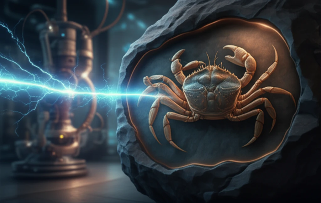
Neutron imaging reveals hidden details within a fossil crab.

Neutron Imaging: Unlocking Hidden Details in Life Sciences

"Explore how neutron imaging is revolutionizing research in paleontology, soil science, and dentistry."


Imagine a world where we could peer inside delicate biological samples without causing any harm, revealing hidden structures and compositions with incredible precision. This is the promise of neutron imaging, a cutting-edge technique that's rapidly transforming research across multiple scientific fields. Unlike traditional methods like X-rays or MRIs, neutron imaging offers a unique way to visualize the internal features of samples without compromising their integrity.

Neutron imaging works by using neutrons, subatomic particles that interact differently with matter than X-rays. Neutrons are particularly sensitive to hydrogen, making them ideal for imaging biological specimens rich in water and organic material. As neutrons pass through a sample, they are scattered and absorbed to varying degrees depending on the material they encounter. By detecting the neutrons that emerge, scientists can create detailed images of the sample's internal structure.

While neutron imaging has been around for some time, recent advancements in neutron sources and detector systems have made it more accessible and powerful than ever before. The IMAT (Imaging and Materials Science & Engineering) beamline at the ISIS Neutron and Muon Source in the UK is at the forefront of this revolution, offering researchers a state-of-the-art facility to explore the potential of neutron imaging in a wide range of applications. Let's dive into some exciting examples of how neutron imaging is unlocking hidden details in paleontology, soil science, and dentistry.

Why Neutron Imaging is a Game-Changer for Paleontology

Neutron imaging reveals hidden details within a fossil crab.

Paleontology, the study of prehistoric life, often relies on techniques that can be destructive or provide limited information. X-ray computed tomography (CT) is a common tool, but it struggles when the fossil and the surrounding rock have similar compositions. This is where neutron tomography shines. Because neutrons interact differently with materials, they can distinguish between the fossil and the matrix, even when X-rays fail.

Consider the case of fossil crabs from the Eocene period in Spain. X-ray CT scans provided limited detail, but neutron tomography at IMAT revealed previously unseen features within the fossils. This breakthrough allows paleontologists to study these ancient creatures in unprecedented detail, gaining new insights into their anatomy and evolution.

  • Non-destructive Analysis: Preserves the integrity of rare and valuable fossils.
  • Enhanced Contrast: Distinguishes between fossil and surrounding rock, even with similar densities.
  • Internal Feature Visualization: Reveals hidden details of fossilized structures.
This ability to see the unseeable makes neutron tomography an invaluable tool for paleontologists, complementing traditional methods and opening new avenues for research. By providing a clearer picture of fossilized remains, neutron imaging helps us understand the history of life on Earth.

The Future of Life Science Research

Neutron imaging is more than just a scientific tool; it's a gateway to new discoveries and a deeper understanding of the world around us. As technology advances and access to facilities like IMAT expands, we can expect to see even more groundbreaking applications of neutron imaging in the years to come. From unraveling the mysteries of ancient life to optimizing agricultural practices and improving dental health, neutron imaging is poised to revolutionize life science research and beyond.

About this Article -

This article was crafted using a human-AI hybrid and collaborative approach. AI assisted our team with initial drafting, research insights, identifying key questions, and image generation. Our human editors guided topic selection, defined the angle, structured the content, ensured factual accuracy and relevance, refined the tone, and conducted thorough editing to deliver helpful, high-quality information.See our About page for more information.

Everything You Need To Know

1

What is neutron imaging and why is it considered so valuable for studying biological samples?

Neutron imaging is a technique that uses neutrons to visualize the internal structure of samples non-destructively. It's particularly useful for biological specimens because neutrons are highly sensitive to hydrogen, which is abundant in water and organic materials. This allows scientists to see details that might be invisible to other imaging methods, such as X-rays or MRIs. The significance of neutron imaging lies in its ability to provide unique insights without damaging the sample, making it invaluable for fields like paleontology, soil science, and dentistry where preserving the integrity of the specimen is crucial.

2

What is neutron tomography and how is it particularly useful in the field of paleontology?

Neutron tomography is a specific application of neutron imaging used to create 3D reconstructions of a sample's internal structure. It's especially valuable in paleontology because it can differentiate between a fossil and the surrounding rock matrix, even when X-rays can't. This enhanced contrast allows paleontologists to visualize hidden details within fossils, providing new information about their anatomy and evolution. The implications are significant, as neutron tomography enables non-destructive analysis of rare and valuable fossils, leading to a deeper understanding of prehistoric life.

3

What is the IMAT beamline, and why is it important for advancing neutron imaging research?

The IMAT (Imaging and Materials Science & Engineering) beamline is a state-of-the-art facility at the ISIS Neutron and Muon Source in the UK. It's important because it provides researchers with access to advanced neutron imaging capabilities, allowing them to explore a wide range of applications. IMAT's advanced technology facilitates groundbreaking research in fields like paleontology, soil science, and dentistry. The availability of such facilities is crucial for advancing neutron imaging techniques and expanding their use in various scientific disciplines.

4

How is neutron imaging being used in paleontology, and what are the benefits of using this technique in this field?

In paleontology, neutron imaging is used to study fossils without damaging them. Neutrons can distinguish between the fossil and the surrounding rock, revealing hidden details that X-rays might miss. This is significant because it allows paleontologists to gain new insights into the anatomy and evolution of ancient creatures, such as fossil crabs from the Eocene period. The implications include a better understanding of the history of life on Earth and the preservation of rare and valuable fossils for future research.

5

Besides paleontology, soil science, and dentistry, what other potential applications exist for neutron imaging?

While the text highlights applications in paleontology, soil science, and dentistry, neutron imaging has potential uses in various other fields. For example, in materials science, it can be used to study the internal structure of materials and components without causing damage. In agriculture, neutron imaging can help optimize irrigation strategies by visualizing water distribution in soil and plants. These broader applications underscore the versatility and importance of neutron imaging as a scientific tool, paving the way for future discoveries and innovations.

Newsletter Subscribe

Subscribe to get the latest articles and insights directly in your inbox.