Detailed ultrasound image of a parathyroid gland

Neck Ultrasound: Your Unsung Hero in Parathyroid Health?

"Discover how this non-invasive tool can precisely locate parathyroid adenomas, guide surgical decisions, and potentially minimize invasive procedures."


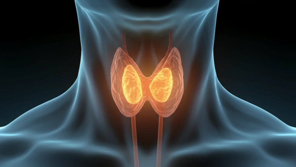
For individuals facing primary hyperparathyroidism (PHPT), parathyroidectomy offers a promising path to recovery. This surgical procedure, honed over decades, has evolved significantly with the advent of precise preoperative localization techniques.

Initially, standard bilateral neck exploration was the norm. Now, minimally invasive parathyroidectomy is increasingly favored. This shift is largely due to our enhanced ability to accurately locate parathyroid adenomas (PTAs) before surgery.

Ultrasonography (US) has emerged as a cornerstone in this evolution. It's non-invasive, relatively inexpensive, and quicker compared to other imaging methods. This article delves into the accuracy and utility of neck ultrasounds in identifying and characterizing parathyroid adenomas.

Why Neck Ultrasounds Are a Game-Changer for Parathyroid Adenomas

Detailed ultrasound image of a parathyroid gland

A study published in Otolaryngology-Head and Neck Surgery evaluated the effectiveness of ultrasonography (US) in estimating both the size and location of parathyroid adenomas (PTAs). The study involved 410 patients who underwent parathyroidectomy for primary hyperparathyroidism between 1996 and 2012.

The results indicated that US correctly localized the adenoma in 76% of cases, demonstrating a sensitivity of 76.2% and a positive predictive value of 86.8%. However, the measurements were least accurate for adenomas smaller than 1 cm in diameter, with only 24% being correctly identified.

  • High Accuracy: Correctly identifies adenomas in a majority of cases.
  • Non-Invasive: Provides detailed imaging without invasive procedures.
  • Operator-Dependent: Accuracy significantly improves with experienced specialists.
  • Size Matters: More effective for larger adenomas (over 1 cm).
The study also highlighted the impact of operator expertise. Scans performed by a single, specialized senior operator had a significantly higher accuracy rate than those conducted by multiple operators, particularly for small adenomas (P < .001).

The Future of Parathyroid Care: Precision Through Ultrasound

Neck ultrasounds are invaluable for evaluating parathyroid adenomas. They offer precision in determining size and location, but expertise matters, especially for smaller adenomas. As technology and expertise advance, expect even greater accuracy, ensuring better outcomes for individuals undergoing parathyroid treatment. Prioritize experienced specialists for the most reliable results.

About this Article -

This article was crafted using a human-AI hybrid and collaborative approach. AI assisted our team with initial drafting, research insights, identifying key questions, and image generation. Our human editors guided topic selection, defined the angle, structured the content, ensured factual accuracy and relevance, refined the tone, and conducted thorough editing to deliver helpful, high-quality information.See our About page for more information.

This article is based on research published under:

DOI-LINK: 10.1177/0194599818792236, Alternate LINK

Title: Accuracy Of Neck Ultrasonography In Predicting The Size And Location Of Parathyroid Adenomas

Subject: Otorhinolaryngology

Journal: Otolaryngology–Head and Neck Surgery

Publisher: SAGE Publications

Authors: Sagit Stern, Sharon Tzelnick, Aviram Mizrachi, Maya Cohen, Thomas Shpitzer, Gideon Bachar

Published: 2018-08-14

Everything You Need To Know

1

What is a neck ultrasound, and why is it important for parathyroid health?

A neck ultrasound is a non-invasive imaging technique used to visualize the parathyroid glands in the neck. It's crucial because it helps locate parathyroid adenomas, which are benign tumors that can cause primary hyperparathyroidism. This is important because precise localization of these adenomas allows for more targeted surgical removal, improving patient outcomes and potentially minimizing the extent of surgery needed.

2

What exactly is a parathyroid adenoma, and why is it important to identify it?

A parathyroid adenoma (PTA) is a benign tumor that forms on one or more of the parathyroid glands. These glands regulate calcium levels in the blood. PTAs can cause the glands to overproduce parathyroid hormone (PTH), leading to primary hyperparathyroidism (PHPT). Identifying the location and size of a parathyroid adenoma is crucial for planning surgical removal, known as a parathyroidectomy, and restoring normal calcium levels.

3

What is ultrasonography, and what role does it play in relation to parathyroid adenomas?

Ultrasonography (US) is significant because it is a non-invasive and relatively inexpensive imaging method used to locate parathyroid adenomas (PTAs) before surgery. The use of US is important because US enables surgeons to perform minimally invasive parathyroidectomy instead of standard bilateral neck exploration. US is also quicker compared to other imaging methods, but its accuracy is dependent on the size of the adenoma and the experience of the operator.

4

What is primary hyperparathyroidism, and how does it relate to parathyroid adenomas and neck ultrasounds?

Primary hyperparathyroidism (PHPT) is a condition where one or more of the parathyroid glands become overactive and produce too much parathyroid hormone (PTH), leading to elevated calcium levels in the blood. This can cause various health problems. If PHPT is caused by a parathyroid adenoma, the recommended treatment is typically a parathyroidectomy to remove the adenoma and restore normal parathyroid function and calcium levels. Neck ultrasounds play a critical role in locating these adenomas prior to surgery.

5

What is a minimally invasive parathyroidectomy, and how does it depend on neck ultrasounds?

Minimally invasive parathyroidectomy is a surgical procedure used to remove parathyroid adenomas (PTAs) through a small incision, guided by preoperative imaging such as neck ultrasonography (US). This approach is favored over standard bilateral neck exploration because it is less invasive, results in smaller scars, and often leads to quicker recovery times for patients. Accurate localization of PTAs using neck ultrasounds is essential for the success of minimally invasive parathyroidectomy.

Newsletter Subscribe

Subscribe to get the latest articles and insights directly in your inbox.