Illustration of a jugular vein aneurysm in the human neck

Neck Swelling? Understanding and Managing Jugular Vein Aneurysms

"Learn about jugular vein aneurysms, including causes, symptoms, diagnosis, and treatment options for this rare condition."


Venous aneurysms, particularly those affecting the head and neck region, are rare. A saccular aneurysm of the external jugular vein with thrombus is exceptionally uncommon. This article explores a case of saccular aneurysm of the external jugular vein with partial thrombosis in a 30-year-old woman, diagnosed and treated by surgical excision.

Jugular venous aneurysms and phlebectasias are rare venous malformations. Phlebectasia typically presents as a fusiform dilatation without tortuosity, often congenital and seen in childhood. In contrast, venous aneurysms, often saccular, usually occur in adulthood, frequently secondary to trauma or vein-related diseases.

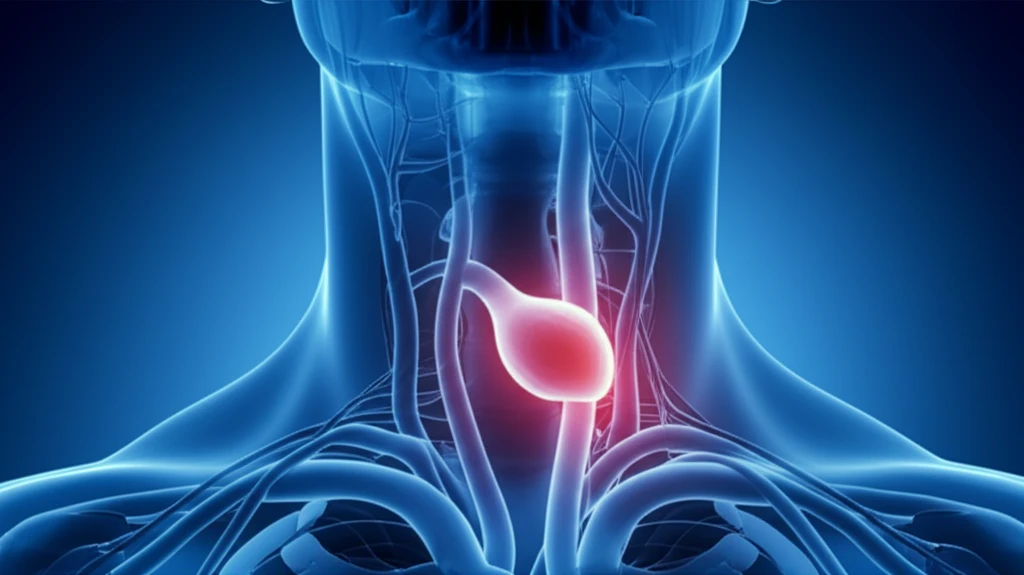
Jugular venous aneurysms typically manifest as soft, compressible, painless neck swellings that enlarge with straining or the Valsalva maneuver. Accurate diagnosis requires differentiating them from other cystic neck swellings that also increase in size under strain, often using imaging technologies such as ultrasound with color Doppler, computed tomography, or magnetic resonance imaging.

What are the Symptoms and Diagnosis of Jugular Vein Aneurysms?

Illustration of a jugular vein aneurysm in the human neck

A key symptom of a jugular venous aneurysm is a soft, compressible swelling in the neck. This swelling often becomes more prominent when straining or performing the Valsalva maneuver, which involves attempting to exhale against a closed airway. It's important to note that these aneurysms are generally painless.

To confirm a diagnosis, healthcare providers use various imaging techniques:

  • Ultrasound with Color Doppler: This is often the first step, as it can show the structure of the vein and blood flow.
  • Computed Tomography (CT): Provides detailed cross-sectional images of the neck.
  • Magnetic Resonance Imaging (MRI): Offers even more detailed views of soft tissues and blood vessels.
These imaging methods help doctors distinguish venous aneurysms from other conditions, such as cysts or tumors in the neck. The images can reveal the size and shape of the aneurysm, as well as any complications like thrombosis (clotting).

When is Surgery Necessary for Jugular Vein Aneurysms?

While jugular phlebectasia is often managed conservatively, surgical excision is typically recommended for saccular aneurysms to prevent potential complications such as thrombosis or thromboembolism. Surgery involves removing the affected section of the vein, a procedure that has proven effective in resolving symptoms and preventing future issues. If you notice unusual swelling in your neck, prompt medical evaluation can lead to an accurate diagnosis and appropriate treatment, ensuring peace of mind and long-term health.

About this Article -

This article was crafted using a human-AI hybrid and collaborative approach. AI assisted our team with initial drafting, research insights, identifying key questions, and image generation. Our human editors guided topic selection, defined the angle, structured the content, ensured factual accuracy and relevance, refined the tone, and conducted thorough editing to deliver helpful, high-quality information.See our About page for more information.

This article is based on research published under:

DOI-LINK: 10.1177/0218492316686477, Alternate LINK

Title: Saccular Aneurysm Of External Jugular Vein With Partial Thrombosis

Subject: Cardiology and Cardiovascular Medicine

Journal: Asian Cardiovascular and Thoracic Annals

Publisher: SAGE Publications

Authors: Hrishikesh Sukhadeo Parashi, Kunal Hemant Rawekar, Manoj Murlidhar Joshi, Hemant Shivram Namdev, Manish Ramdhan Jadhao, Krishnarao Narayan Bhosle

Published: 2016-12-20

Everything You Need To Know

1

What are the telltale signs and how is a jugular vein aneurysm diagnosed?

A jugular venous aneurysm is characterized by a soft, compressible swelling in the neck that often becomes more noticeable when straining or performing the Valsalva maneuver. This swelling is typically painless. Diagnosis involves imaging techniques such as ultrasound with color Doppler, computed tomography (CT), or magnetic resonance imaging (MRI) to differentiate it from other conditions like cysts or tumors in the neck.

2

Under what circumstances would surgery be deemed necessary for a jugular vein aneurysm?

Surgical excision is often recommended for saccular aneurysms. This is because of the risk of complications such as thrombosis (clotting) or thromboembolism. Surgical removal of the affected vein section helps to resolve symptoms and prevent potential future issues.

3

How do jugular venous aneurysms differ from jugular phlebectasias, and why is this distinction important?

Jugular venous aneurysms are often saccular and usually occur in adulthood, frequently resulting from trauma or vein-related diseases. In contrast, jugular phlebectasias usually appear as fusiform dilatations without any twisting; they are typically congenital and are found during childhood.

4

What role do imaging techniques play in diagnosing jugular vein aneurysms?

Ultrasound with color Doppler is important because it is often the first step in diagnosing jugular venous aneurysms. This method allows healthcare providers to visualize the structure of the vein and assess blood flow. Computed Tomography (CT) and Magnetic Resonance Imaging (MRI) offer more detailed cross-sectional views of the neck and soft tissues. The imaging techniques help differentiate venous aneurysms from other conditions and determine the size and shape of the aneurysm, and any presence of complications like thrombosis.

5

How common is a saccular aneurysm of the external jugular vein with thrombus, and why is it significant?

A saccular aneurysm of the external jugular vein with thrombus is an exceptionally uncommon occurrence. This is important because it requires careful diagnosis and management due to the potential for complications. Understanding the causes, symptoms, and diagnostic methods is essential for healthcare providers to effectively address this rare condition through surgical excision.

Newsletter Subscribe

Subscribe to get the latest articles and insights directly in your inbox.