Nail Perineurioma: Spotting the Rare Tumor with Sonography
"Understand how sonography aids in diagnosing nail perineurioma, a rare benign tumor affecting the nail structure, and what this means for early detection and treatment."
Have you noticed an unusual change in your nail's appearance, such as swelling or dystrophy? While many nail conditions are common, some rare cases require specialized attention. One such condition is nail perineurioma, a benign tumor that affects the neural tissue around the nail. Though rare, understanding this condition and how it's diagnosed is crucial for early and effective management.
Nail perineurioma is a benign tumor derived from neural tissue and is exceptionally rare, particularly when it occurs in the nail area. Unlike more common nerve tumors like schwannomas or neurofibromas, perineuriomas originate from perineurial cells, which surround nerve fascicles. Identifying this specific type of tumor is essential for proper diagnosis and treatment.
The symptoms of nail perineurioma can often mimic other common nail conditions, making diagnosis challenging. Swelling, clubbing, or dystrophy of the nail are typical signs, which could easily be mistaken for fibromas or exostosis. Therefore, advanced diagnostic tools like sonography play a vital role in accurately identifying nail perineurioma.
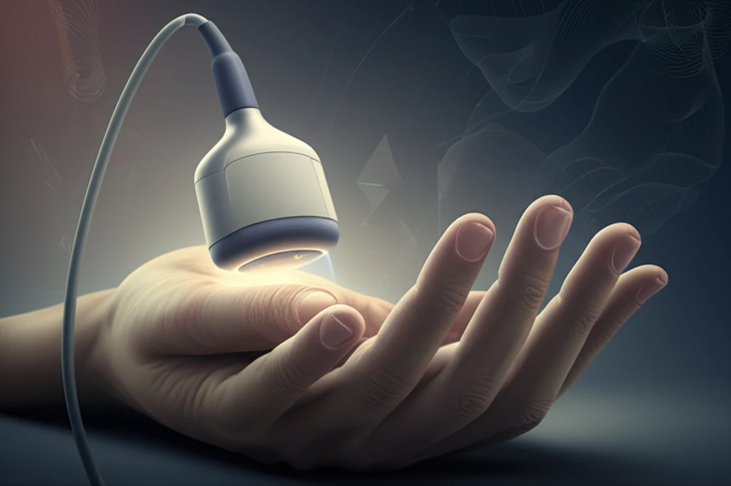
The Role of Sonography in Diagnosing Nail Perineurioma
Sonography, or ultrasound imaging, has emerged as a valuable tool in diagnosing nail perineurioma. Traditional methods may not always capture the subtle nuances of this rare tumor, but sonography provides a non-invasive way to visualize the affected area in detail. Using high-frequency sound waves, sonography can reveal the tumor's size, location, and characteristics, aiding in accurate diagnosis.
- A poorly defined hypoechoic mass on the radial aspect of the nail bed.
- Involvement of the ungual matrix and extension into the lateral nail fold.
- Measurements of the tumor's dimensions.
- Minimal blood flow within the mass, as shown by Color Doppler sonography.
The Future of Nail Perineurioma Diagnosis
Sonography offers a non-invasive and reliable method for diagnosing nail perineuriomas, supporting accurate and timely interventions. As technology advances, we can expect even more precise and detailed imaging techniques to improve diagnostic accuracy. This will lead to better patient outcomes and a higher quality of life for those affected by this rare condition. If you notice any unusual changes in your nails, consult with a healthcare professional to explore the possibilities of sonography and other advanced diagnostic methods.