Digital illustration depicting the liver with glowing cysts, symbolizing intra-peritoneal deposits, and a blurred image of an ovarian cyst removal, highlighting the medical history.

Mysteries of the Ovary: Unraveling the Rare Case of Intra-peritoneal Deposits

"From Surgery to Surprise: A Look into a Rare Complication and What it Means for Women's Health"


In the realm of women's health, the complexities of the female reproductive system often present intriguing medical cases. Among these, ovarian cysts are a common occurrence, but when they lead to unexpected complications, they offer a fascinating insight into the body's intricate workings. This article delves into a rare medical scenario: intra-peritoneal deposits resulting from a previously resected ovarian mature benign cystic teratoma, commonly known as a dermoid cyst. This exploration aims to shed light on the unusual progression of this condition, its diagnosis, and the surgical interventions required, emphasizing the importance of understanding such rare occurrences in medical practice.

Ovarian cysts, while typically benign, can sometimes lead to unforeseen complications. This case highlights a situation where surgical intervention, meant to resolve an ovarian cyst, inadvertently led to the seeding of cystic deposits within the peritoneal cavity. The term 'intra-peritoneal' refers to the space within the abdomen that houses the organs, and the presence of these deposits raises questions about the behavior of these cysts and the body's response to them. This article will investigate the diagnostic processes, surgical procedures, and the implications of such findings, providing an informative look into the less common aspects of women's health.

This article is designed to provide a clear, concise understanding of a complex medical situation. It will navigate through the symptoms, diagnostic methods, and surgical treatments involved in managing intra-peritoneal deposits from a mature benign cystic teratoma. The goal is to present this information in an accessible manner, offering insights for both medical professionals and individuals interested in learning more about women's health and the nuances of medical conditions. The focus is on simplifying complex medical concepts, making them understandable and relevant to a broader audience.

Unveiling the Intrigue: The Case of Intra-peritoneal Deposits

Digital illustration depicting the liver with glowing cysts, symbolizing intra-peritoneal deposits, and a blurred image of an ovarian cyst removal, highlighting the medical history.

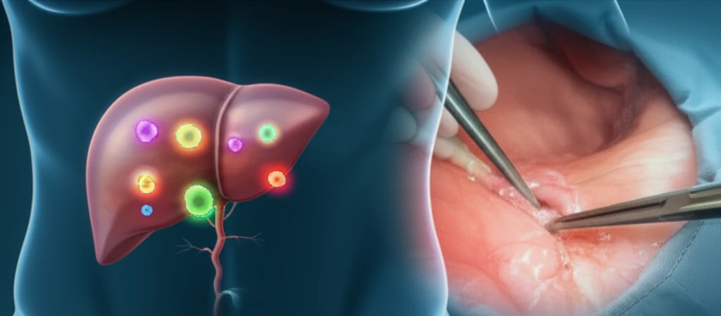
The medical case begins with a patient who presented with persistent upper abdominal pain, leading to a series of investigative steps. Initial findings from an ultrasound revealed three hypoechoic, septated masses in the right upper quadrant of the abdomen. Further investigation using a contrast-enhanced CT scan provided more detailed insights into the nature of these lesions. These scans revealed the presence of multiple mixed-density lesions along the liver surface, which contained fat, soft tissue, and calcification, suggesting a complex composition. The medical team carefully reviewed the patient's previous imaging history, including a CT scan from five years prior, which showed no such lesions but indicated the presence of a 10 cm ovarian mature cystic teratoma. This historical context was crucial in piecing together the puzzle.

Additional imaging, specifically an MR liver scan, showed that these lesions were deposited along the liver's surface, particularly in the right subdiaphragmatic region, close to the inferior vena cava. The MR scans displayed high signal intensity on both T1 and T2 weighted images, indicating the presence of macroscopic fat. This observation, coupled with the chemical shift artifact and suppression on the short tau inversion recovery (STIR) sequences, further confirmed the presence of fat within the lesions. These diagnostic findings pointed towards the possibility of intra-peritoneal deposits, raising questions about their origin and nature.

  • Initial Ultrasound Findings: Revealed multiple masses in the right upper quadrant.
  • CT Scan Insights: Showed mixed-density lesions with fat, soft tissue, and calcification.
  • Historical Context: Review of previous scans pointed to a history of an ovarian mature cystic teratoma.
  • MR Liver Scan: Highlighted fat-containing lesions along the liver surface.
  • Diagnostic Conclusions: Led to the possibility of intra-peritoneal deposits related to the ovarian teratoma.
The diagnostic process, which involved a combination of ultrasound, CT scans, and MRI, along with the patient's surgical history, led to a specific radiological diagnosis. The medical team suspected intra-peritoneal mature benign cystic teratomas, originating from seeding during the earlier resection of the ovarian mature benign cystic teratoma. This surgical complication is not commonly documented, emphasizing the importance of recognizing and understanding this rare occurrence. The case was discussed in a hepatobiliary multidisciplinary team meeting, demonstrating a collaborative approach to patient care and diagnostic precision.

Key Takeaways and Further Insights

This case underscores the significance of comprehensive medical evaluation, particularly in cases where a patient presents with unusual symptoms and a history of previous procedures. Understanding the potential for complications, like intra-peritoneal deposits, requires a collaborative approach involving multiple diagnostic methods and a keen understanding of the patient's medical history. This case provides essential insights into recognizing and managing a rare complication, thereby contributing to improved patient care and a deeper understanding of women's health. Continued research and documentation of such cases are vital to enhancing our medical knowledge and providing the best possible outcomes for patients.

About this Article -

This article was crafted using a human-AI hybrid and collaborative approach. AI assisted our team with initial drafting, research insights, identifying key questions, and image generation. Our human editors guided topic selection, defined the angle, structured the content, ensured factual accuracy and relevance, refined the tone, and conducted thorough editing to deliver helpful, high-quality information.See our About page for more information.

Everything You Need To Know

1

What are the key initial findings that led to the investigation of intra-peritoneal deposits in this medical case?

The investigation began with a patient experiencing persistent upper abdominal pain. Initial findings from an ultrasound revealed three hypoechoic, septated masses in the right upper quadrant of the abdomen. Further investigation with a contrast-enhanced CT scan showed multiple mixed-density lesions along the liver surface, containing fat, soft tissue, and calcification. These findings prompted a deeper look into the patient's medical history, revealing a previous ovarian mature benign cystic teratoma, which was crucial in understanding the progression of the condition.

2

How did the medical team determine the nature of the lesions found along the liver surface, and what specific imaging techniques were used?

The medical team utilized a combination of imaging techniques to determine the nature of the lesions. Following the initial ultrasound and CT scan, an MR liver scan was performed. This scan highlighted fat-containing lesions deposited along the liver surface, particularly in the right subdiaphragmatic region, near the inferior vena cava. The MR scans showed high signal intensity on both T1 and T2 weighted images, indicating macroscopic fat. Further confirmation came from the presence of a chemical shift artifact and suppression on the short tau inversion recovery (STIR) sequences, which are specific indicators of fat within the lesions. These findings were critical in suggesting the presence of intra-peritoneal deposits.

3

What is the significance of a "mature benign cystic teratoma" in relation to the intra-peritoneal deposits described, and why is this case considered rare?

The ovarian mature benign cystic teratoma, also known as a dermoid cyst, is a type of ovarian cyst that can contain various tissues, such as hair, teeth, and fat. In this case, the intra-peritoneal deposits were suspected to have originated from seeding during a prior resection of a mature benign cystic teratoma. This means that during the surgical removal of the original cyst, some of its contents may have spilled into the peritoneal cavity, leading to the formation of new deposits. The rarity of this occurrence highlights the unexpected nature of this surgical complication, underscoring the importance of thorough surgical techniques and awareness of potential complications in women's health.

4

What diagnostic steps were crucial in diagnosing intra-peritoneal deposits, and how did the patient's history play a role?

The diagnosis of intra-peritoneal deposits relied on a combination of imaging studies and a thorough review of the patient's medical history. Initial diagnostic steps included an ultrasound, which revealed suspicious masses, followed by CT scans and MRIs. The CT scans provided insights into the composition of the lesions, while the MRIs confirmed the presence of fat. However, the patient's surgical history of having a mature benign cystic teratoma was critical in linking the current findings to a potential cause. The medical team connected the dots by comparing the current imaging results with the history of the ovarian cyst resection, leading them to suspect that the deposits were related to seeding during the earlier surgery.

5

Why is it important for medical professionals to understand and recognize rare complications like intra-peritoneal deposits from mature benign cystic teratomas, and what is the value of multidisciplinary team meetings in this context?

Understanding and recognizing rare complications, such as intra-peritoneal deposits from a mature benign cystic teratoma, is crucial for improving patient care and outcomes. These rare cases underscore the importance of comprehensive medical evaluation, which involves a detailed review of the patient's history and the use of various diagnostic methods. In this case, the collaborative approach of a hepatobiliary multidisciplinary team meeting was essential. This allowed for multiple specialists to contribute their expertise, leading to a more precise diagnosis and informing the best course of treatment. Such collaboration ensures that all aspects of the case are considered, leading to improved patient outcomes and enhancing overall understanding of women's health complications.

Newsletter Subscribe

Subscribe to get the latest articles and insights directly in your inbox.