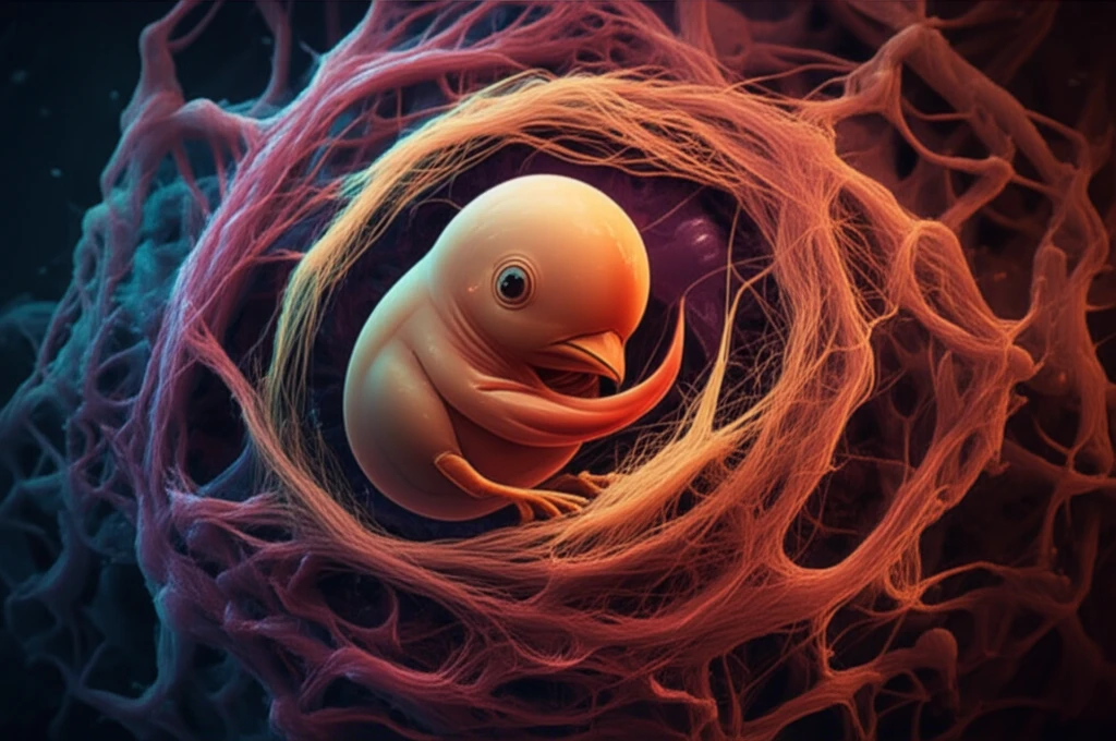
Chick embryo cells intertwined with muscle fibers, symbolizing muscle dystrophy research.

Muscle Dystrophy Research: Can Chick Embryo Cells Lead to New Treatments?

"Scientists are exploring chick embryo cells to create models for studying muscle dystrophy and testing new therapies, offering hope for a cost-effective approach to understanding and treating this challenging disease."


Muscle dystrophy (MD) is a group of genetic diseases characterized by progressive muscle weakness and degeneration. These conditions arise from defects in proteins crucial for muscle structure and function. While there is currently no cure for MD, scientists are continuously searching for ways to better understand the disease and develop effective treatments.

Traditional research models, such as stem cells and genetically modified mice, present challenges like ethical concerns, high costs, and complex genetic manipulations. Researchers are now exploring alternative models that are more accessible and can provide valuable insights into the disease mechanisms.

A recent study published in In Vitro Cellular & Developmental Biology - Animal investigates the potential of using chick embryo cells as an in vitro model for studying muscle dystrophy. This innovative approach offers a cost-effective and ethically sound method for understanding the disease and testing potential therapies.

Chick Embryo Cells: A Novel Approach to Muscle Dystrophy Research

Chick embryo cells intertwined with muscle fibers, symbolizing muscle dystrophy research.

The study, conducted by Verma Urja, Kashmira Khaire, Suresh Balakrishnan, and Gowri Kumari Uggini, focuses on utilizing chick embryo cells to mimic the characteristics of muscle dystrophy in a laboratory setting. The researchers isolated limb myoblasts – precursor cells to muscle fibers – from 11-day-old chick embryos. These cells were then cultured and treated with an anti-dystroglycan antibody (IIH6).

The IIH6 antibody targets alpha-dystroglycan, a protein essential for connecting the cytoskeleton within muscle cells to the extracellular matrix. This connection is vital for maintaining muscle integrity and function. Disrupting this link is a key feature of muscle dystrophy.

  • Mimicking Muscle Dystrophy: By blocking the function of alpha-dystroglycan with the IIH6 antibody, the researchers aimed to replicate the cellular characteristics of muscle dystrophy in the cultured chick embryo cells.
  • Observing Cellular Changes: The scientists then observed the treated muscle cells, looking for changes in their morphology (structure), contractibility (ability to contract), and gene expression (the activity of specific genes).
  • Analyzing Results: These observations were compared to control cultures to determine the impact of the alpha-dystroglycan blockade.
The results of the study showed promising evidence that chick embryo cells can serve as a viable model for studying muscle dystrophy. The IIH6-treated muscle cells exhibited several key features associated with the disease, including:

The Future of Muscle Dystrophy Research

This study suggests that chick embryo cells offer a valuable, accessible tool for studying muscle dystrophy and potentially identifying new therapeutic targets. This model provides a cost-effective alternative to traditional methods, making it easier for researchers to investigate the disease mechanisms and test potential treatments. Further research is needed to fully explore the potential of this model and translate these findings into clinical applications for individuals affected by muscle dystrophy.

About this Article -

This article was crafted using a human-AI hybrid and collaborative approach. AI assisted our team with initial drafting, research insights, identifying key questions, and image generation. Our human editors guided topic selection, defined the angle, structured the content, ensured factual accuracy and relevance, refined the tone, and conducted thorough editing to deliver helpful, high-quality information.See our About page for more information.

Everything You Need To Know

1

What exactly is muscle dystrophy, and why is it important to study?

Muscle dystrophy (MD) refers to a group of genetic diseases that cause progressive weakening and degeneration of muscles. These conditions stem from defects in proteins vital for muscle structure and function. Although there is currently no cure, ongoing research strives to understand the disease better and develop effective treatments. The significance of understanding MD lies in the potential to alleviate the suffering of those affected and improve their quality of life.

2

What are chick embryo cells, and how are they being used in muscle dystrophy research?

Chick embryo cells are being explored as a novel model for studying muscle dystrophy in a laboratory setting. Researchers isolate limb myoblasts, which are precursor cells to muscle fibers, from chick embryos. These cells are then cultured and treated to mimic the characteristics of muscle dystrophy, allowing scientists to observe cellular changes and test potential therapies in a cost-effective and ethically sound manner.

3

What is the role of the IIH6 antibody in studying muscle dystrophy, and why is it important?

The IIH6 antibody is significant in muscle dystrophy research because it targets alpha-dystroglycan, a protein crucial for connecting the cytoskeleton within muscle cells to the extracellular matrix. By blocking the function of alpha-dystroglycan with the IIH6 antibody, researchers can replicate the cellular characteristics of muscle dystrophy in cultured chick embryo cells. Disrupting the connection of the cytoskeleton to the extracellular matrix is a key feature of muscle dystrophy.

4

What are limb myoblasts and why are they used in this type of research?

Limb myoblasts are precursor cells to muscle fibers, playing a crucial role in muscle development and regeneration. In the context of muscle dystrophy research using chick embryo cells, limb myoblasts are isolated from chick embryos and cultured to mimic the characteristics of muscle dystrophy in a laboratory setting. Studying these cells allows researchers to observe cellular changes and test potential therapies, offering insights into the disease mechanisms.

5

What is the extracellular matrix, and what role does it play in muscle dystrophy?

The extracellular matrix is essential for providing structural support and biochemical cues to surrounding cells, including muscle cells. It plays a crucial role in maintaining muscle integrity and function. In the context of muscle dystrophy, disruptions in the connection between the cytoskeleton within muscle cells and the extracellular matrix can lead to muscle weakness and degeneration. The extracellular matrix provides the environment surrounding the cells.

Newsletter Subscribe

Subscribe to get the latest articles and insights directly in your inbox.