Muscle Dystrophy Breakthrough: Chick Embryo Cells Offer New Hope for In Vitro Modeling
"Researchers are leveraging chick embryonic cells to create an accessible and cost-effective in vitro model for studying muscle dystrophy, potentially paving the way for novel pharmacological agents."
For decades, scientists have been meticulously studying the molecular mechanisms governing muscle development in vertebrates. Key players like Myoblast Determination Protein 1 (MYOD1), myogenin (MYOG), Myogenic Factor 5 (MYF5), and Myogenic Regulatory Factor 4 (MRF4) have been identified as critical myogenic regulatory factors since the late 1980s. These factors orchestrate muscle formation during embryonic development, as well as maintenance and regeneration processes post-embryonic development.
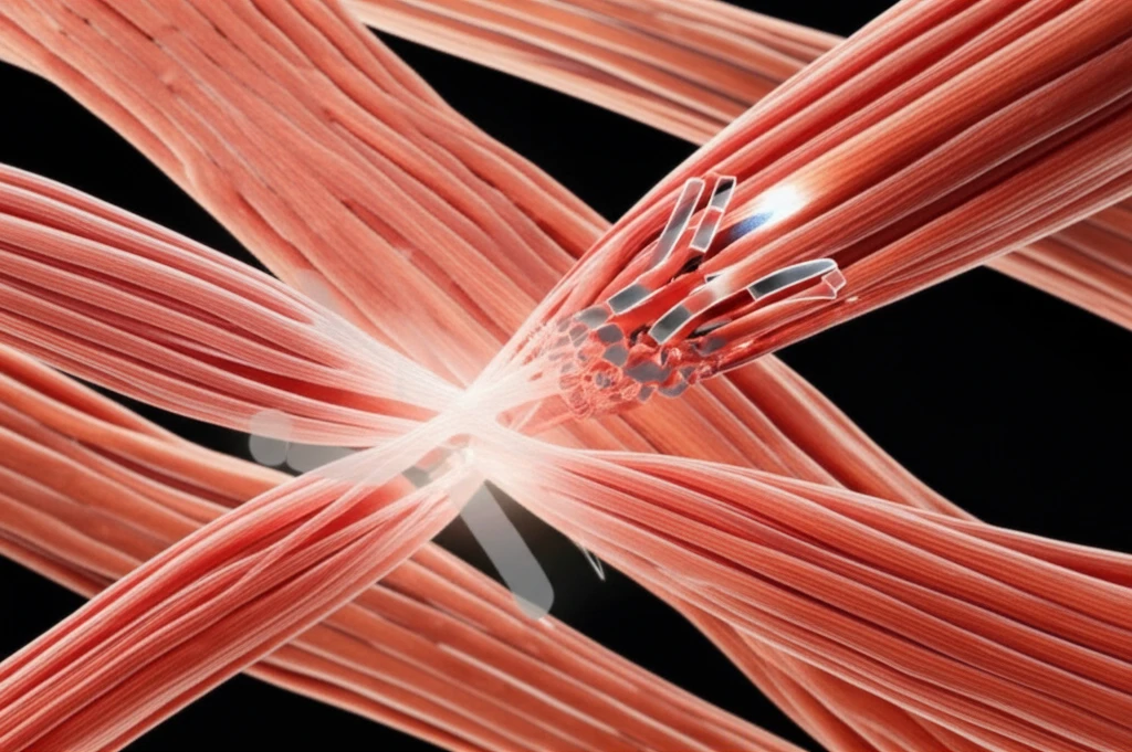
The vertebrate sarcolemma, which is the cell membrane of muscle fibers, contains a sophisticated assembly of glycoproteins. These include alpha and beta subtypes of dystroglycan, as well as alpha, beta, and gamma subtypes of sarcoglycan and dystrophin. When isolated, these components were found to be closely associated with the dystrophin protein, giving rise to the term 'dystrophin-glycoprotein complex.' Genetic defects in any of these proteins can lead to severe muscle developmental issues, with muscular dystrophy (MD) being a prime example.
Muscular dystrophy (MD) is a devastating disease characterized by progressive muscle weakness and atrophy. Among the various forms of MD, Duchenne muscular dystrophy (DMD) is the most prevalent. DMD arises from genetic mutations—such as deletions, insertions, point mutations, or duplications—affecting the dystrophin gene. Researchers are constantly seeking effective treatments, and novel models are needed to better understand the disease.
Why Chick Embryonic Cells?
Chick embryonic cells offer a compelling alternative for in vitro MD studies, addressing many limitations associated with stem cells and induced myogenic cell lines. The conventional methods often grapple with ethical concerns, extended timelines for myogenic cell derivation, genetic modifications during cell induction, high costs, and limited cell yields. The myogenesis process in chicks closely mirrors that in humans, making them a relevant model. Significant early research on myogenesis in limbs and other organs was conducted using chick embryos. Their ease of manipulation, cost-effectiveness, and considerable genetic similarity to mammals make them an invaluable asset.
- Cell Isolation and Culture: Limb myoblasts from 11-day-old chick embryos were isolated and cultured.
- MD Induction: The cultures were treated with IIH6 antibody to disrupt the connection between cytoskeletal proteins and the extracellular matrix.
- Observation: Changes in morphometrics, contractibility, and mRNA expression were monitored.
The Future of MD Research
This innovative approach using chick embryonic cells provides a simple and cost-effective method to further understand the disease mechanism and conduct initial studies on the effects of novel pharmacological agents. By offering a readily accessible and ethically sound model, researchers can accelerate the development of effective treatments for muscular dystrophy, bringing new hope to those affected by this debilitating condition.