PET/CT scan highlighting cancerous activity in bones.

Multiple Myeloma: How PET/CT Scans are Revolutionizing Diagnosis and Care

"Discover how Positron Emission Tomography (PET) combined with Computed Tomography (CT) is transforming the landscape of multiple myeloma management, offering new hope for early detection and personalized treatment strategies."


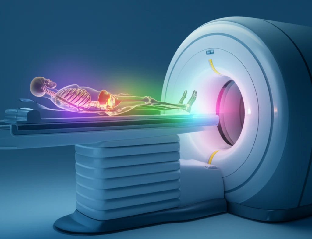
In the ongoing battle against multiple myeloma (MM), a cancer of plasma cells, new imaging techniques are providing revolutionary advancements in how the disease is diagnosed and managed. Historically, the assessment of bone disease in MM, a critical factor in determining the disease's severity and progression, relied on conventional radiology. However, the introduction of Positron Emission Tomography (PET) combined with Computed Tomography (CT), known as PET/CT, has significantly altered the landscape. This advanced imaging technique offers a more sensitive and comprehensive approach to detecting bone lesions and assessing the extent of the disease.

The importance of accurate and early detection of bone disease in MM cannot be overstated. The presence and severity of bone lesions are key criteria in defining active or symptomatic MM, influencing treatment decisions and overall patient management. PET/CT imaging provides a functional view of the body, highlighting areas of increased metabolic activity, which can indicate the presence of cancerous cells even before they are visible on traditional X-rays. This capability is particularly valuable in identifying unsuspected disease sites, both within the bones and in extra-medullary locations, offering a more complete picture of the disease burden.

This article delves into the transformative role of PET/CT scans in multiple myeloma, examining how this technology is enhancing diagnostic accuracy, improving risk stratification, and guiding treatment strategies. We will explore how PET/CT is being used to refine the criteria for diagnosing MM, assess treatment response, and predict patient outcomes, ultimately contributing to more personalized and effective care for individuals living with this challenging disease.

Why PET/CT is a Game-Changer in Multiple Myeloma

PET/CT scan highlighting cancerous activity in bones.

PET/CT's superiority over traditional methods lies in its ability to provide both anatomical and functional information. While CT scans excel at visualizing the structural details of bones, PET scans detect metabolic activity, revealing areas where cancer cells are actively growing. By integrating these two technologies, PET/CT offers a comprehensive assessment of bone disease, identifying lesions that might be missed by conventional radiology.

The International Myeloma Working Group has recognized the importance of PET/CT by incorporating it into the diagnostic criteria for multiple myeloma. The presence of osteolytic bone destruction, identified through skeletal radiography, CT, or PET/CT, now fulfills the criteria for bone disease in MM. This inclusion underscores the growing recognition of PET/CT as a valuable tool for early and accurate diagnosis.

  • Enhanced Sensitivity: PET/CT detects metabolically active lesions that may not be visible on X-rays.
  • Comprehensive Assessment: Combines anatomical and functional information for a complete picture of the disease.
  • Improved Accuracy: Helps differentiate between benign and malignant bone lesions.
  • Early Detection: Identifies bone disease at an earlier stage, allowing for prompt treatment.
PET/CT imaging plays a critical role in assessing the extent and pattern of bone disease in multiple myeloma. It can highlight the number and intensity of bone lesions, differentiate between diffuse and focal involvement, and identify extra-medullary disease (EMD). The presence of EMD and a higher number of focal lesions are associated with a poorer prognosis, providing valuable information for risk stratification and treatment planning. The standardized uptake value (SUV), a measure of metabolic activity, has also been shown to have prognostic significance in some studies.

The Future of Multiple Myeloma Care with PET/CT

While PET/CT is not a perfect imaging technique for MM, its increasing use is allowing us to better understand the behavior of MM-related bone disease and to take appropriate action at the right time, thus contributing to improve the care of our patients. As we continue to unravel the full potential of PET/CT, we can expect even more refined diagnostic and treatment strategies, ultimately improving the lives of individuals affected by multiple myeloma.

About this Article -

This article was crafted using a human-AI hybrid and collaborative approach. AI assisted our team with initial drafting, research insights, identifying key questions, and image generation. Our human editors guided topic selection, defined the angle, structured the content, ensured factual accuracy and relevance, refined the tone, and conducted thorough editing to deliver helpful, high-quality information.See our About page for more information.

Everything You Need To Know

1

What is Multiple Myeloma, and why is early detection so crucial in managing this disease?

Multiple Myeloma (MM) is a cancer of plasma cells, impacting the bone marrow and often leading to bone lesions. Early detection is paramount because the presence and severity of bone lesions directly influence treatment decisions and overall patient management. Advanced imaging techniques like PET/CT scans allow for earlier and more accurate identification of these lesions, leading to prompt intervention and potentially better outcomes for individuals affected by MM.

2

How does PET/CT differ from traditional methods like conventional radiology in diagnosing Multiple Myeloma?

Traditional methods, such as conventional radiology, primarily focus on the structural details of bones. PET/CT, however, offers a more comprehensive approach. It integrates Positron Emission Tomography (PET) and Computed Tomography (CT). CT scans provide detailed anatomical information, while PET scans detect metabolic activity. This combined approach allows for the identification of metabolically active lesions, which might be missed by conventional radiology, offering a more complete picture of the disease burden in Multiple Myeloma.

3

How does the International Myeloma Working Group incorporate PET/CT into the diagnosis of Multiple Myeloma, and what does this signify?

The International Myeloma Working Group has incorporated PET/CT into the diagnostic criteria for Multiple Myeloma. The presence of osteolytic bone destruction, identified through skeletal radiography, CT, or PET/CT, now fulfills the criteria for bone disease in MM. This inclusion signifies the growing recognition of PET/CT as a valuable tool for early and accurate diagnosis, reflecting its ability to detect disease that may not be visible through traditional methods.

4

What specific advantages does PET/CT offer in assessing the extent and pattern of bone disease in Multiple Myeloma?

PET/CT provides a detailed view of bone disease in Multiple Myeloma by highlighting the number and intensity of bone lesions. It can differentiate between diffuse and focal involvement, as well as identify extra-medullary disease (EMD). The presence of EMD and a higher number of focal lesions are associated with a poorer prognosis. Furthermore, the standardized uptake value (SUV), a measure of metabolic activity, has also been shown to have prognostic significance, offering valuable information for risk stratification and treatment planning.

5

What is the future role of PET/CT in Multiple Myeloma care, and what impact can we expect it to have on patients?

The increasing use of PET/CT is allowing for a better understanding of the behavior of Multiple Myeloma-related bone disease. As we continue to unravel the full potential of PET/CT, we can expect even more refined diagnostic and treatment strategies. This may lead to more personalized and effective care for individuals with Multiple Myeloma. This includes earlier detection, improved risk stratification, and the ability to monitor treatment response, ultimately leading to improved patient outcomes and quality of life.

Newsletter Subscribe

Subscribe to get the latest articles and insights directly in your inbox.