AI correcting motion in a PET scan of a heart.

Motion Artifacts in PET Scans: How AI Can Help

"New research reveals how data-driven AI can detect and compensate for patient movement during coronary PET imaging, leading to more accurate diagnoses."


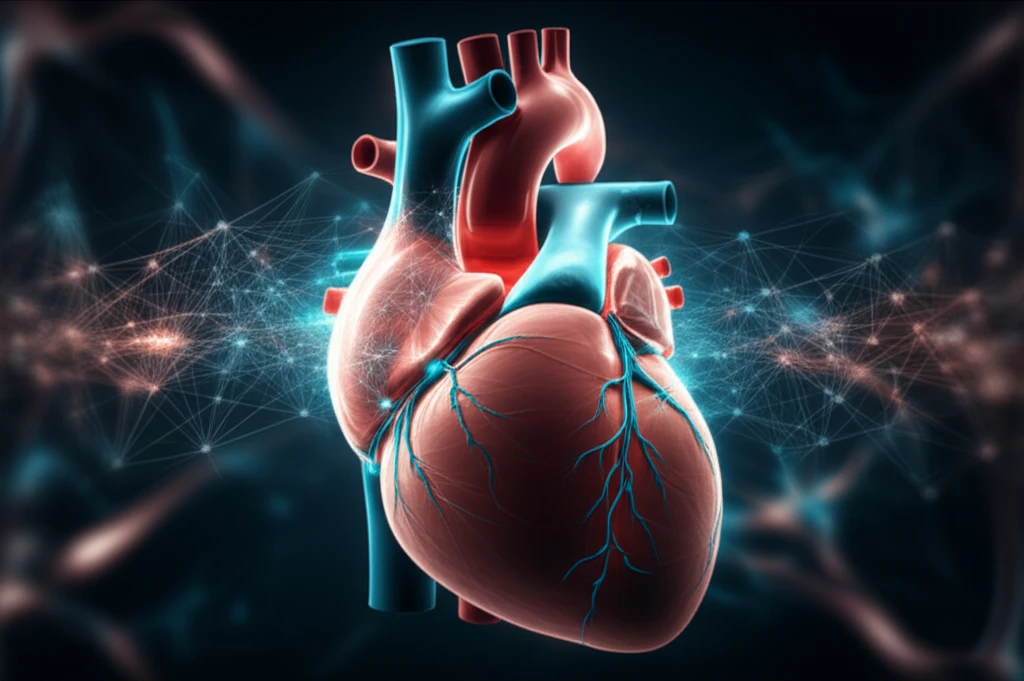
Medical imaging, particularly Positron Emission Tomography (PET), relies on patients remaining still for extended periods. However, even slight movements can blur images, creating artifacts that compromise the accuracy of diagnoses. This is especially critical in coronary imaging, where precise detection of micro-calcification is crucial.

Coronary PET imaging often requires scans lasting up to 30 minutes, increasing the likelihood of patient movement. These movements, referred to as gross patient motion (GPM), can significantly degrade image quality and affect quantitative assessments. Consequently, there is a growing need for methods that can automatically detect and compensate for GPM during PET scans.

A recent study published in the Journal of Nuclear Medicine investigates a novel data-driven approach for detecting and correcting patient motion during coronary 18F-NaF PET imaging. This method leverages artificial intelligence to analyze data and compensate for movement, leading to more accurate and reliable results. Let's explore this technology and what it means for the future of cardiac PET imaging.

AI to the Rescue: How Motion Detection Works

AI correcting motion in a PET scan of a heart.

The cornerstone of this innovative approach is a data-driven algorithm designed to track patient movement by monitoring the center-of-mass (CoM) of count rates within the PET data. This technique analyzes data every 200 milliseconds, enabling it to capture even subtle shifts in patient position. The study focused on two primary types of patient motion:

  • Sudden Repositioning (SR): Abrupt movements exceeding 0.5 mm within 3 seconds.
  • General Repositioning (GR): Gradual drifts greater than 0.3 mm over 15 seconds or more.

Sudden Repositioning (SR): Abrupt movements exceeding 0.5 mm within 3 seconds. General Repositioning (GR): Gradual drifts greater than 0.3 mm over 15 seconds or more.
After identifying instances of GPM, the algorithm reconstructs diastolic images and co-registers individual GPM frames with focal coronary uptake in 3D. This process creates a GPM-compensated (GPMC) image series, essentially correcting for the movement that occurred during the scan.

The Future of Cardiac PET Imaging

The study's findings suggest that GPM is a common occurrence during coronary 18F-NaF PET imaging and can significantly affect the accuracy of quantitative assessments. By implementing an automated motion compensation technique, clinicians can potentially improve diagnostic accuracy and reduce the risk of misdiagnosis.

This AI-driven approach offers a practical solution for addressing motion-related artifacts in PET imaging. It doesn't require any additional hardware or modifications to existing scanning protocols, making it easily adaptable to current clinical workflows. The retrospective nature of the method means it can also be applied to previously acquired data, unlocking new insights from past studies.

As AI continues to evolve, its applications in medical imaging will likely expand, leading to more precise, efficient, and personalized healthcare. This study represents a significant step toward harnessing the power of AI to improve the accuracy and reliability of cardiac PET imaging, ultimately benefiting patients and clinicians alike.

About this Article -

This article was crafted using a human-AI hybrid and collaborative approach. AI assisted our team with initial drafting, research insights, identifying key questions, and image generation. Our human editors guided topic selection, defined the angle, structured the content, ensured factual accuracy and relevance, refined the tone, and conducted thorough editing to deliver helpful, high-quality information.See our About page for more information.

This article is based on research published under:

DOI-LINK: 10.2967/jnumed.118.217877, Alternate LINK

Title: Data-Driven Gross Patient Motion Detection And Compensation: Implications For Coronary 18F-Naf Pet Imaging

Subject: Radiology, Nuclear Medicine and imaging

Journal: Journal of Nuclear Medicine

Publisher: Society of Nuclear Medicine

Authors: Martin Lyngby Lassen, Jacek Kwiecinski, Sebastien Cadet, Damini Dey, Chengjia Wang, Marc R. Dweck, Daniel S. Berman, Guido Germano, David E. Newby, Piotr J. Slomka

Published: 2018-11-15

Everything You Need To Know

1

What are the main problems that patient movement causes during PET scans?

Patient movement, often referred to as Gross Patient Motion (GPM), is a significant concern in Positron Emission Tomography (PET) scans. Even small movements can blur images, creating artifacts that compromise the accuracy of diagnoses, particularly in coronary imaging. Specifically, Sudden Repositioning (SR) and General Repositioning (GR) are two types of motion that can affect image quality. The impact is critical because it can lead to misdiagnoses due to inaccurate image interpretation.

2

How exactly does the new AI approach detect and correct patient movement?

The data-driven AI approach works by tracking patient movement using an algorithm that monitors the center-of-mass (CoM) of count rates within the PET data. This algorithm analyzes data every 200 milliseconds to detect subtle shifts in patient position. This analysis allows the AI to identify instances of Gross Patient Motion (GPM), including Sudden Repositioning (SR) and General Repositioning (GR). The algorithm then reconstructs diastolic images and co-registers individual GPM frames to create a GPM-compensated (GPMC) image series, correcting for the movement that occurred during the scan. The main concept here is the AI's ability to analyze and correct for patient movement, thereby improving image quality.

3

Why is patient motion especially problematic in coronary PET imaging?

Coronary PET imaging is particularly susceptible to motion artifacts because it often requires scans lasting up to 30 minutes. This extended duration increases the likelihood of Gross Patient Motion (GPM). Moreover, precise detection of micro-calcification is crucial in coronary imaging for accurate diagnoses, and even minor movements can blur the images, making accurate diagnosis difficult. The new AI method allows for more reliable results, as it can correct for the effects of Sudden Repositioning (SR) and General Repositioning (GR).

4

What are the different types of patient movement addressed by the AI?

The two primary types of patient motion are Sudden Repositioning (SR) and General Repositioning (GR). Sudden Repositioning (SR) refers to abrupt movements exceeding 0.5 mm within 3 seconds, while General Repositioning (GR) involves gradual drifts greater than 0.3 mm over 15 seconds or more. These types of Gross Patient Motion (GPM) can degrade image quality and affect quantitative assessments. The AI algorithm is designed to identify and compensate for both SR and GR, improving the accuracy of the scan.

5

What is the ultimate impact of using AI to correct motion artifacts in PET scans?

By implementing an automated motion compensation technique, the potential impact of Gross Patient Motion (GPM) can be mitigated, improving diagnostic accuracy. This automated process using AI can correct for both Sudden Repositioning (SR) and General Repositioning (GR) and has the potential to reduce the risk of misdiagnosis. This is especially important for coronary 18F-NaF PET imaging, where accurate detection is crucial. The implications for patients and clinicians involve more reliable diagnoses and potentially fewer unnecessary interventions.

Newsletter Subscribe

Subscribe to get the latest articles and insights directly in your inbox.