Comparative visualization of mixed dentition analysis using X-ray and CBCT imaging.

Mixed Dentition Analysis: Is Advanced Imaging Worth the Cost?

"Cone-beam computed tomography (CBCT) offers precision in assessing developing teeth, but does it outweigh traditional methods and added expense?"


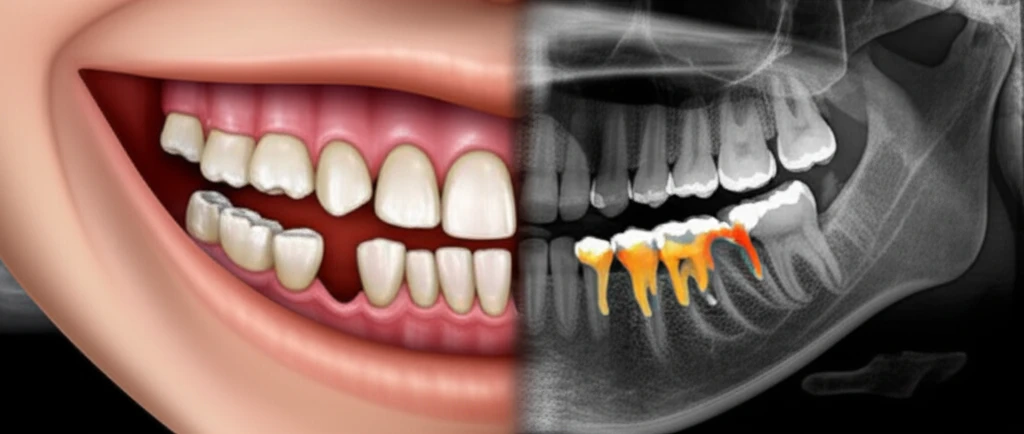
Achieving a harmonious balance between tooth size and jaw dimensions is a cornerstone of successful orthodontic treatment. Malocclusions often stem from discrepancies between these factors. The mixed dentition phase, a period where both primary and permanent teeth coexist, presents unique challenges in assessing potential crowding and developing a comprehensive treatment plan.

During this transitional period, slight crowding in the lower arch is often considered normal as the erupting permanent lateral incisors require additional space. However, accurately predicting the size of unerupted permanent teeth becomes crucial to determine whether the dental arch can accommodate them without significant crowding or the need for extensive intervention.

Traditional methods for mixed dentition analysis include clinical examination, study models, and radiographic assessments. While these techniques have served orthodontists for decades, the advent of cone-beam computed tomography (CBCT) has opened new avenues for visualizing and measuring dental structures with greater precision. This article delves into a study comparing CBCT with traditional methods, evaluating the accuracy, advantages, and limitations of each approach.

CBCT vs. Traditional Radiography: A Comparative Look at Mixed Dentition Analysis

Comparative visualization of mixed dentition analysis using X-ray and CBCT imaging.

The study featured in the Dental Press Journal of Orthodontics aimed to evaluate the effectiveness of CBCT in assessing the size of intra-osseous teeth during the mixed dentition phase. The researchers compared CBCT measurements with those obtained using traditional methods, including:

  • Moyers’ Analysis: A prediction table based on the mesiodistal width of erupted lower incisors to estimate the size of unerupted canines and premolars.
  • Tanaka-Johnston Formula: A similar predictive method that uses a formula instead of a table.
  • 45-Degree Oblique Radiographs: A radiographic technique used to visualize unerupted teeth and measure their dimensions.

  • Plaster Cast Models: Measurements of erupted incisors were taken on plaster models using digital calipers.
  • CBCT Scans: The same dental units were gauged by means of Dolphin software resources.
  • Statistical Analysis: High agreement was observed between tomographic and radiographic methods.
  • Low Agreement: Between tomographs and other methods being evaluated.
The study revealed a high level of agreement between CBCT and 45-degree oblique radiographs. However, there was less agreement between CBCT measurements and the predictions derived from Moyers' analysis and the Tanaka-Johnston formula. This suggests that while CBCT and oblique radiographs provide comparable assessments, predictive methods may not always accurately reflect individual tooth dimensions.

The Verdict: Is CBCT the Future of Mixed Dentition Analysis?

While CBCT offers several advantages, including the ability to visualize intra-osseous teeth individually and without superimposition of anatomical structures, it is essential to consider the limitations and costs associated with this technology. CBCT scans expose patients to higher radiation doses than traditional radiographs, and the cost of CBCT equipment and software can be significant. Ultimately, the decision to use CBCT for mixed dentition analysis should be based on a careful assessment of the individual patient's needs, the complexity of the case, and the available resources.

About this Article -

This article was crafted using a human-AI hybrid and collaborative approach. AI assisted our team with initial drafting, research insights, identifying key questions, and image generation. Our human editors guided topic selection, defined the angle, structured the content, ensured factual accuracy and relevance, refined the tone, and conducted thorough editing to deliver helpful, high-quality information.See our About page for more information.

Everything You Need To Know

1

What is mixed dentition analysis and why is it important?

Mixed dentition analysis aims to achieve a harmonious balance between tooth size and jaw dimensions. It is significant because malocclusions often stem from discrepancies between these factors. During the mixed dentition phase, where both primary and permanent teeth are present, accurately predicting the size of unerupted permanent teeth is crucial to determine whether the dental arch can accommodate them without significant crowding or extensive intervention. Without this analysis, orthodontic treatments might not be as effective or could lead to unnecessary procedures.

2

What are the traditional methods used for mixed dentition analysis?

Traditional methods for mixed dentition analysis include clinical examination, study models, and radiographic assessments. Methods such as Moyers’ Analysis, the Tanaka-Johnston Formula, and 45-degree oblique radiographs have been used. While these techniques have served orthodontists for decades, they each have limitations in visualizing and measuring dental structures, especially unerupted teeth. They may not always accurately reflect individual tooth dimensions, potentially leading to less precise treatment planning.

3

What is cone-beam computed tomography and how is it used in mixed dentition analysis?

Cone-beam computed tomography, or CBCT, offers a way to visualize and measure dental structures with greater precision compared to traditional methods. It allows orthodontists to see intra-osseous teeth individually and without the superimposition of anatomical structures. CBCT can provide a more accurate assessment of unerupted teeth. However, it also exposes patients to higher radiation doses and involves significant costs for equipment and software.

4

What are Moyers’ Analysis and the Tanaka-Johnston Formula, and why are they significant?

Moyers’ Analysis is a prediction table based on the mesiodistal width of erupted lower incisors used to estimate the size of unerupted canines and premolars. The Tanaka-Johnston Formula is a similar predictive method that uses a formula instead of a table to achieve the same goal. These methods are important because they provide a way to estimate the size of unerupted teeth using measurements from erupted teeth. However, these predictive methods may not always accurately reflect individual tooth dimensions, as highlighted by comparisons with CBCT measurements.

5

How should a dentist decide whether to use cone-beam computed tomography for mixed dentition analysis?

The decision to use cone-beam computed tomography for mixed dentition analysis should be based on a careful assessment of the individual patient's needs, the complexity of the case, and the available resources. CBCT scans expose patients to higher radiation doses than traditional radiographs, and the cost of CBCT equipment and software can be significant. If the case is straightforward and traditional methods provide sufficient information, CBCT may not be necessary. However, in complex cases where precise visualization of unerupted teeth is crucial, the benefits of CBCT may outweigh the risks and costs.

Newsletter Subscribe

Subscribe to get the latest articles and insights directly in your inbox.