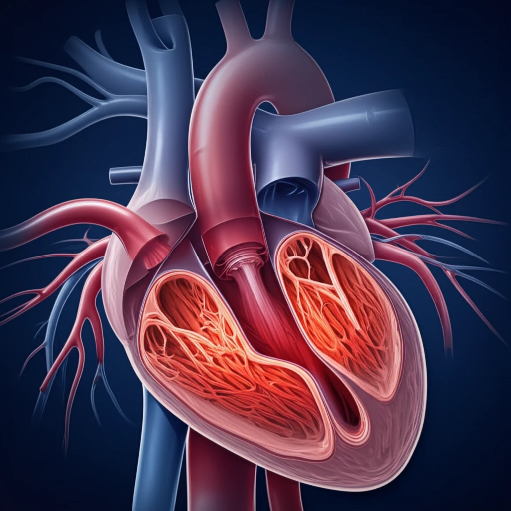
Illustration of a heart with an enlarged aortic root, representing the connection between mitochondrial dysfunction and aortic root ectasia.

Mitochondrial Disorders: How Aortic Root Ectasia Can Be a Key Indicator

"Unveiling the link between aortic root ectasia and mitochondrial dysfunction for earlier diagnosis and improved management."


Mitochondrial disorders (MIDs) are a complex group of conditions that affect how the body produces energy. These disorders can manifest in many ways, impacting various organs and systems. While some symptoms are well-recognized, others are rarer, making diagnosis challenging.

One such rare manifestation is arteriopathy, a disease of the arteries. Arteriopathy in MIDs can take several forms, including atherosclerosis, stenosis, aneurysm, and ectasia. Aortic root ectasia (ARE), characterized by an enlarged aorta near the heart, has recently emerged as a potential indicator of underlying mitochondrial dysfunction.

This article explores the connection between ARE and MIDs, drawing on a recent case study that highlights the importance of considering mitochondrial dysfunction in patients presenting with unexplained ARE. By understanding this link, healthcare professionals can improve diagnostic accuracy and potentially offer earlier interventions.

Decoding the Connection: Aortic Root Ectasia and Mitochondrial Disorders

Illustration of a heart with an enlarged aortic root, representing the connection between mitochondrial dysfunction and aortic root ectasia.

Aortic root ectasia (ARE) is defined as an aortic diameter of 40-50 mm at the level of the aortic valves. While ARE can result from various factors, including arterial hypertension, Marfan syndrome, and congenital heart defects, its association with mitochondrial disorders is increasingly recognized.

Mitochondrial disorders (MIDs) are genetic conditions affecting the mitochondria, the powerhouses of our cells. These disorders impair energy production and can affect multiple organ systems, leading to a wide range of symptoms. The connection between MIDs and ARE lies in the potential impact of mitochondrial dysfunction on vascular smooth muscle cells (VSMCs).

  • VSMCs are crucial for maintaining the structure and function of the aorta.
  • Mitochondrial dysfunction can impair VSMC function, leading to weakening and dilation of the aortic root.
  • ARE, in the context of a MID, may indicate that the metabolic defect is affecting the vascular system.
A recent case study illustrates this connection. An 84-year-old woman with a history of various health issues, including dementia, hypothyroidism, and atrial fibrillation, was found to have ARE. Further investigation revealed other signs suggestive of a mitochondrial disorder, including elevated lactate levels and abnormalities on brain imaging. While a definitive diagnosis of MID could not be established before her death, the case highlights the importance of considering mitochondrial dysfunction in patients with unexplained ARE and multiple system involvement.

Looking Ahead: Implications for Diagnosis and Management

The recognition of ARE as a potential feature of MIDs has important implications for diagnosis and management. Patients presenting with unexplained ARE, particularly those with other signs suggestive of mitochondrial dysfunction, should be evaluated for possible underlying MIDs. This evaluation may involve a thorough medical history, physical examination, blood tests (including lactate levels), and imaging studies. While genetic testing can help confirm the diagnosis, it's not always conclusive, and a high index of suspicion is crucial. Long-term monitoring is essential for patients with ARE and MIDs to detect any progression of aortic dilation and prevent potential complications, such as aortic dissection or rupture.

About this Article -

This article was crafted using a human-AI hybrid and collaborative approach. AI assisted our team with initial drafting, research insights, identifying key questions, and image generation. Our human editors guided topic selection, defined the angle, structured the content, ensured factual accuracy and relevance, refined the tone, and conducted thorough editing to deliver helpful, high-quality information.See our About page for more information.

Everything You Need To Know

1

What is Aortic Root Ectasia (ARE), and what does it signify?

Aortic Root Ectasia (ARE) is defined as an aortic diameter of 40-50 mm at the level of the aortic valves. It signifies an enlargement of the aorta near the heart. While ARE can stem from conditions like arterial hypertension or congenital heart defects, its presence can also be a key indicator of underlying mitochondrial disorders (MIDs). The enlargement suggests a weakening of the aortic wall, potentially due to factors like impaired function of Vascular Smooth Muscle Cells (VSMCs).

2

How are Mitochondrial Disorders (MIDs) linked to Aortic Root Ectasia (ARE)?

The connection between Mitochondrial Disorders (MIDs) and Aortic Root Ectasia (ARE) lies in the impact of mitochondrial dysfunction on Vascular Smooth Muscle Cells (VSMCs). MIDs, which are genetic conditions, impair energy production within the mitochondria. This dysfunction can affect the VSMCs, crucial for maintaining the aorta's structure and function. Impaired VSMC function leads to weakening and dilation of the aortic root, resulting in ARE. Thus, ARE, in the context of a MID, may indicate that the metabolic defect is affecting the vascular system.

3

Besides ARE, what other signs might suggest a Mitochondrial Disorder (MID)?

Besides Aortic Root Ectasia (ARE), other signs suggestive of a Mitochondrial Disorder (MID) can include various symptoms affecting multiple organ systems. Elevated lactate levels in blood tests are a common indicator of mitochondrial dysfunction. Brain imaging may reveal abnormalities, indicating neurological involvement. Given that MIDs can manifest in many ways, a wide range of symptoms impacting different organs and systems can be indicative. Diagnosis often requires considering a combination of these factors.

4

How can healthcare professionals use the connection between Aortic Root Ectasia (ARE) and Mitochondrial Disorders (MIDs) to improve patient care?

Healthcare professionals can leverage the connection between Aortic Root Ectasia (ARE) and Mitochondrial Disorders (MIDs) to improve diagnostic accuracy and facilitate earlier intervention. By recognizing ARE as a potential feature of MIDs, clinicians can evaluate patients with unexplained ARE, particularly those showing other signs indicative of mitochondrial dysfunction, for possible underlying MIDs. This includes detailed medical history, physical examination, blood tests (e.g., lactate levels), and imaging studies. This approach enables early detection, allowing for interventions to manage the progression of the disorder and prevent complications.

5

What steps are involved in evaluating and managing a patient with Aortic Root Ectasia (ARE) suspected of having a Mitochondrial Disorder (MID)?

Evaluating a patient with Aortic Root Ectasia (ARE) suspected of having a Mitochondrial Disorder (MID) involves several steps. Initially, a thorough medical history is taken, and a physical examination is conducted to assess the patient's overall health and identify any relevant symptoms. Blood tests, including lactate levels, are crucial for identifying metabolic abnormalities. Imaging studies, such as echocardiograms and potentially MRIs, are used to assess the aorta and other affected organs. While genetic testing can help confirm the diagnosis, it is not always conclusive. Long-term monitoring is essential for patients with ARE and MIDs to detect any progression of aortic dilation and prevent potential complications such as aortic dissection or rupture. Early diagnosis and comprehensive management are key to improving patient outcomes.

Newsletter Subscribe

Subscribe to get the latest articles and insights directly in your inbox.