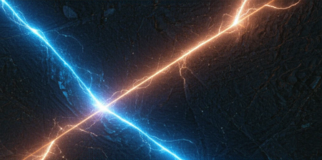
Microscopic alpha particle tracks glowing against a mineral background.

Micron-Scale Mapping: How Autoradiography Unlocks Secrets of Radioactive Minerals

"Explore the innovative technique of alpha particle autoradiography and its groundbreaking potential for high-resolution radionuclide mapping in environmental science and mineralogy."


The ability to pinpoint the exact location of radioactive elements within materials is crucial across various fields, from safely managing mining operations to ensuring the long-term security of nuclear waste storage. Traditional methods often fall short when dealing with low-activity samples, where radionuclides are sparsely distributed and challenging to detect. This is especially true for isotopes with short half-lives, which decay rapidly and exist in minuscule concentrations.

Imagine trying to find a single grain of sand on a vast beach – that’s the challenge scientists face when searching for these elusive radioactive hotspots. Current analytical tools lack the sensitivity and spatial precision needed to map these elements at the microscopic level, hindering our understanding of their behavior and potential risks.

Now, a groundbreaking technique is changing the game. Alpha particle autoradiography is emerging as a powerful solution for visualizing and mapping radionuclides with unprecedented resolution. This method allows researchers to directly image the alpha particles emitted during radioactive decay, revealing the precise location of these elements within a material, even at incredibly low concentrations.

What is Alpha Particle Autoradiography and How Does It Work?

Microscopic alpha particle tracks glowing against a mineral background.

At its core, alpha particle autoradiography is like taking a snapshot of radioactive decay. The process involves applying a special emulsion to a sample, much like coating photographic film. This emulsion contains silver halide grains that are sensitive to radiation. When an alpha particle (a positively charged particle emitted during radioactive decay) strikes these grains, it leaves a microscopic track, creating a latent image.

After a carefully controlled exposure period, which can last from days to months depending on the sample's activity, the emulsion is developed. This process converts the silver halide grains along the alpha particle tracks into metallic silver, making them visible under a microscope. The resulting image, known as an autoradiograph, reveals the precise locations where radioactive decay occurred within the sample.

  • High Spatial Resolution: See details down to the micrometer scale, revealing tiny radioactive hotspots.
  • Low Concentration Detection: Works even with very small amounts of radioactive material, thanks to long exposure times.
  • Alpha-Specific: Only detects alpha particles, ignoring other types of radiation and reducing background noise.
  • In-Situ Development: Develop the film directly on the sample for the highest possible resolution.
  • Enhanced Contrast: Use polarized light to make the tracks stand out clearly against complex backgrounds.
Think of it like using a specialized camera to capture the faint glow of radioactive decay. This technique opens new doors for understanding the distribution and behavior of radionuclides in a wide range of materials.

The Future of Radionuclide Mapping: Applications and Potential

Alpha particle autoradiography is poised to become an indispensable tool for environmental scientists, mineralogists, and anyone working with radioactive materials. By providing a clear and detailed picture of radionuclide distribution, this technique empowers researchers to address critical challenges in resource management, waste disposal, and environmental protection. The ability to identify and characterize localized areas of high activity unlocks new avenues for understanding the behavior of these elements and mitigating their potential risks. Future research will undoubtedly focus on refining this technique and expanding its applications across diverse scientific and industrial fields.

About this Article -

This article was crafted using a human-AI hybrid and collaborative approach. AI assisted our team with initial drafting, research insights, identifying key questions, and image generation. Our human editors guided topic selection, defined the angle, structured the content, ensured factual accuracy and relevance, refined the tone, and conducted thorough editing to deliver helpful, high-quality information.See our About page for more information.

This article is based on research published under:

DOI-LINK: 10.1016/j.jenvrad.2018.11.008, Alternate LINK

Title: Alpha Particle Autoradiography For High Spatial Resolution Mapping Of Radionuclides

Subject: Health, Toxicology and Mutagenesis

Journal: Journal of Environmental Radioactivity

Publisher: Elsevier BV

Authors: Christopher A.G. Kalnins, Nigel A. Spooner, Michael J.P. Clarke, David Ottaway

Published: 2019-02-01

Everything You Need To Know

1

What is alpha particle autoradiography?

Alpha particle autoradiography is a technique used to visualize and map radionuclides in a material with high resolution. It involves applying a special emulsion containing silver halide grains to a sample. When alpha particles, emitted during radioactive decay, strike these grains, they create microscopic tracks. After development, these tracks become visible as an autoradiograph, revealing the precise locations of radioactive decay.

2

Why is the high resolution of alpha particle autoradiography important?

The high spatial resolution of alpha particle autoradiography allows for the detection of tiny radioactive hotspots at the micrometer scale. This is significant because it enables the examination of low concentrations of radioactive material, which is crucial when dealing with isotopes that have short half-lives and decay rapidly. The technique is also alpha-specific, which reduces background noise by only detecting alpha particles.

3

What are some applications of alpha particle autoradiography in environmental science?

Alpha particle autoradiography can be used to address resource management, waste disposal, and environmental protection challenges. By pinpointing the locations of radionuclides, researchers can better understand the behavior of radioactive elements and mitigate their potential risks. This includes managing mining operations and ensuring the long-term security of nuclear waste storage.

4

Can you explain the basic steps involved in performing alpha particle autoradiography?

The process begins by applying an emulsion containing silver halide grains to a sample. Alpha particles emitted during radioactive decay interact with the emulsion, leaving microscopic tracks. These tracks are then developed into visible metallic silver grains, which reveals the location of the alpha emissions. The autoradiograph can be further enhanced using polarized light for improved contrast.

5

What makes alpha particle autoradiography a powerful technique compared to other methods for detecting radionuclides?

Several features of alpha particle autoradiography contribute to its effectiveness. These include high spatial resolution, low concentration detection, alpha-specificity, in-situ development, and enhanced contrast. Traditional methods often fall short in sensitivity and spatial precision when mapping elements at the microscopic level, hindering the understanding of their behavior and risks.

Newsletter Subscribe

Subscribe to get the latest articles and insights directly in your inbox.