Illustration of lung cancer subtypes, highlighting different blood flow patterns.

Lung Cancer's Hidden Clues: How Blood Flow Reveals the Secrets of AC vs. SCC

"New research unveils the distinct blood flow patterns in adenocarcinoma (AC) and squamous cell carcinoma (SCC), offering crucial insights for personalized lung cancer treatment."


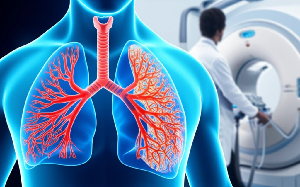
Lung cancer, a formidable adversary, continues to challenge medical science with its complexity and diverse manifestations. Among the various types, adenocarcinoma (AC) and squamous cell carcinoma (SCC) stand out as the two most prevalent non-small cell lung cancer (NSCLC) subtypes. Recent advancements in medical imaging, specifically CT perfusion (CTp), have opened new avenues for understanding the intricate details of these cancers, particularly their blood flow dynamics. This article delves into a groundbreaking study that compares the blood flow characteristics of AC and SCC, offering valuable insights into the disease and its treatment.

The study, published in BioMed Research International, leverages the power of CTp to analyze the blood flow (BF) patterns in AC and SCC tumors. The findings reveal significant differences in how these two subtypes recruit and utilize blood vessels, a critical factor in cancer's growth and response to treatment. By examining these distinct hemodynamic behaviors, researchers are gaining a deeper understanding of the biological mechanisms that drive these cancers and how they might be targeted for improved therapeutic outcomes.

This research not only enhances our knowledge of lung cancer biology but also has the potential to transform clinical practice. The ability to differentiate between AC and SCC based on blood flow patterns could lead to more precise diagnoses, tailored treatment plans, and ultimately, improved patient outcomes. This article will explore the study's methodologies, findings, and implications for the future of lung cancer care, offering a comprehensive overview of this exciting field.

Unveiling the Blood Flow: How CT Perfusion Works

Illustration of lung cancer subtypes, highlighting different blood flow patterns.

CT perfusion is a sophisticated imaging technique that provides detailed information about the blood flow within tissues and tumors. It involves injecting a contrast agent into the patient's bloodstream and then capturing a series of CT scans over a short period. By analyzing how the contrast agent moves through the tumor, radiologists can calculate various perfusion parameters, including blood flow (BF), blood volume, and permeability.

In the context of lung cancer, CTp is used to assess the vascularity of the tumor, which is a measure of the number and function of blood vessels. Tumors, like all tissues, require a blood supply to grow and survive. The more blood vessels a tumor has, the more resources it can access, leading to faster growth and the potential for metastasis. CTp provides a non-invasive way to visualize and quantify these blood vessels, offering valuable insights into the tumor's biology.

  • Contrast Agent Injection: A contrast agent is injected intravenously.
  • Rapid CT Scanning: Multiple CT scans are taken in quick succession.
  • Data Analysis: Specialized software analyzes the movement of the contrast agent.
  • Perfusion Parameters: Blood flow, blood volume, and permeability are calculated.
  • Tumor Assessment: The results help evaluate the tumor's vascularity.
The study in question employed a 256-slice CT system to perform CTp on patients with AC and SCC. The researchers used the maximum-slope method to calculate BF values and implemented techniques to automatically remove unreliable data, ensuring the accuracy of their findings. This meticulous approach allowed them to identify significant differences in the blood flow characteristics of the two cancer subtypes, which we'll explore in detail.

The Future of Lung Cancer Care: Personalized Treatment and Beyond

This study represents a significant step forward in our understanding of lung cancer. By identifying distinct blood flow patterns in AC and SCC, researchers have opened the door to more personalized and effective treatment strategies. As CTp technology continues to evolve, its role in lung cancer diagnosis and management is likely to expand, offering hope for improved outcomes and a brighter future for patients battling this devastating disease.

About this Article -

This article was crafted using a human-AI hybrid and collaborative approach. AI assisted our team with initial drafting, research insights, identifying key questions, and image generation. Our human editors guided topic selection, defined the angle, structured the content, ensured factual accuracy and relevance, refined the tone, and conducted thorough editing to deliver helpful, high-quality information.See our About page for more information.

Everything You Need To Know

1

How does CT perfusion (CTp) work to reveal details about lung tumors?

CT perfusion (CTp) involves injecting a contrast agent into the bloodstream and then taking rapid CT scans. By analyzing how the contrast agent moves through the tumor, radiologists can calculate perfusion parameters such as blood flow (BF), blood volume, and permeability. This helps assess the tumor's vascularity, providing insights into its biology and potential for growth. Currently the technology can differentiate between Adenocarcinoma (AC) and Squamous Cell Carcinoma (SCC). However, it cannot differentiate between all the types of lung cancer. More research and technological advances are needed to expand its capability to cover all other lung cancers.

2

What are adenocarcinoma (AC) and squamous cell carcinoma (SCC), and why is it important to distinguish between them?

Adenocarcinoma (AC) and squamous cell carcinoma (SCC) are two prevalent subtypes of non-small cell lung cancer (NSCLC). Distinguishing between them is crucial because they exhibit different biological behaviors, including variations in blood flow dynamics. These differences can impact how the cancers respond to treatment. Using CT perfusion (CTp) to differentiate between AC and SCC can lead to more precise diagnoses and tailored treatment plans. Currently other forms of NSCLC that are neither AC nor SCC cannot be diagnosed using CTp and require other clinical methods.

3

How might the findings of blood flow differences between adenocarcinoma (AC) and squamous cell carcinoma (SCC) change lung cancer treatment?

Identifying distinct blood flow patterns in adenocarcinoma (AC) and squamous cell carcinoma (SCC) through CT perfusion (CTp) opens the door to more personalized treatment strategies. This understanding allows for more precise diagnoses and the development of tailored treatment plans that target the specific vascular characteristics of each subtype. This tailored approach has the potential to improve treatment outcomes and overall patient care. However, the research focuses solely on AC and SCC, and further investigation is needed to determine whether the understanding of blood flow differences can be expanded to benefit other forms of cancer treatments.

4

What role does blood flow (BF) play in the growth and spread of lung cancer?

Blood flow (BF) is essential for tumor growth and metastasis. Tumors need a blood supply to access resources, and the more blood vessels a tumor has, the faster it can grow and potentially spread. CT perfusion (CTp) helps visualize and quantify these blood vessels, providing valuable insights into the tumor's biology. Differences in BF between adenocarcinoma (AC) and squamous cell carcinoma (SCC) reveal how these subtypes recruit and utilize blood vessels differently, impacting their growth and response to treatment. Although BF is a critical factor, other elements such as the cancer microenvironment and immunological responses can also significantly impact how tumors spread.

5

How does the use of a 256-slice CT system contribute to the accuracy of blood flow (BF) analysis in this type of study?

The use of a 256-slice CT system allows for rapid and detailed imaging during CT perfusion (CTp). This enables researchers to capture the movement of the contrast agent with high precision. Combined with methods to calculate blood flow (BF) values (like the maximum-slope method) and to remove unreliable data, the detailed imaging capability contributes to the accuracy of identifying differences in blood flow characteristics between adenocarcinoma (AC) and squamous cell carcinoma (SCC). However, the 256-slice CT system is only one component of the study, and the reliability of the results also depends on factors such as patient selection, data processing techniques, and the expertise of the radiologists and researchers involved.

Newsletter Subscribe

Subscribe to get the latest articles and insights directly in your inbox.