Lung Cancer Blood Flow: How CT Perfusion Distinguishes Adenocarcinoma and Squamous Cell Carcinoma
"New research reveals that CT perfusion can differentiate between adenocarcinoma and squamous cell carcinoma in lung cancer patients, potentially improving treatment strategies."
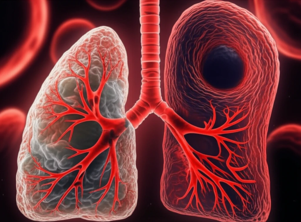
The development of tumors, known as tumorigenesis, relies on angiogenesis—the formation of a complex, often disorganized network of blood vessels that feed the tumor. Understanding these vascular patterns, especially how they change over time, is crucial for characterizing different types of tissue. This is where CT perfusion (CTp) comes in; this advanced imaging technique has gained traction for its ability to provide both high-resolution spatial and temporal data.
CT perfusion works by tracking the journey of a contrast agent as it reaches the tumor, generating time concentration curves (TCCs). By analyzing these curves, doctors can compute perfusion parameters that offer insights into the tumor's blood supply. One particularly valuable parameter is blood flow (BF), which shows a strong link to microvessel density (MVD), a key tissue biomarker. Measuring BF typically involves monitoring the first pass of the contrast medium, which keeps the examination time short and minimizes radiation exposure for the patient.
Using blood flow (BF) information obtained at the time of diagnosis can significantly aid in lesion characterization, particularly for patients who aren't eligible for surgery and need non-surgical treatment plans. What's more, higher baseline BF values in individuals with advanced lung carcinoma may suggest a more favorable response to therapy. The ability to differentiate between responders and non-responders based on BF values highlights the importance of characterizing tumors' hemodynamic profiles, which includes considering different cancer subtypes. By understanding the perfusion characteristics of non-small cell lung cancer (NSCLC), clinicians can gain useful insights into the tumor's status, particularly regarding its degree of hypoxia, which greatly influences how it responds to treatment.
Differentiating Lung Cancer Subtypes: The Role of Blood Flow
Research has indicated that adenocarcinoma (AC) tends to have a significantly lower degree of hypoxia compared to squamous cell carcinoma (SCC). Further studies have explored differences in perfusion parameters among lung cancer subtypes, with some showing conflicting results. To interpret these discrepancies, it’s important to consider potential sources of error in BF computation, such as respiratory motion, CTp artifacts, and tumor location, all of which can affect the reliability of BF values.
- The main aim of the research was to assess and evaluate lung tumors at diagnosis.
- New research was conducted to study the significant differences in perfusion between AC and SCC, the two predominant NSCLC phenotypes.
- To further improve the characterization of AC and SCC perfusion, caused by a high measurement variability, stemming from clinical and physiological factors as well as external causes (e.g., patient movements and artefacts), two methods were used.
Implications for Treatment Planning
This study highlights the importance of understanding blood flow characteristics in different NSCLC subtypes, particularly adenocarcinoma and squamous cell carcinoma. By employing advanced imaging techniques like CT perfusion and carefully analyzing the data to remove artifacts and account for tumor location, clinicians can gain valuable insights into tumor behavior. These insights can inform treatment planning, potentially leading to more effective and personalized approaches, especially with anti-angiogenic therapies.