Resilient Tree of Life: A Symbol of Healing in Lung Cancer Treatment

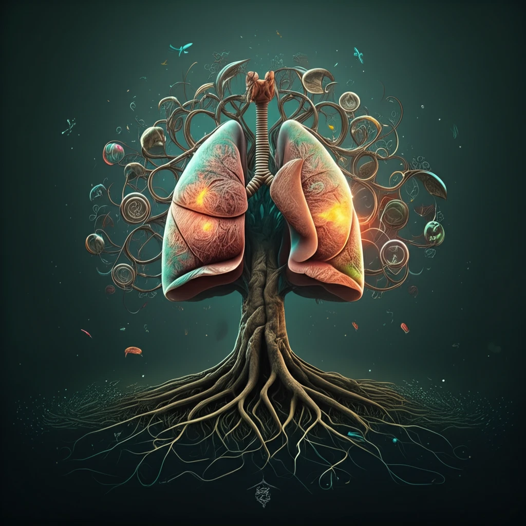
Lung Cancer and Radiotherapy: New Insights into Predicting and Managing Treatment Side Effects

"Discover how cutting-edge radiomics and personalized medicine are transforming lung cancer treatment, offering hope for minimizing radiation-induced complications and improving patient outcomes."


Lung cancer remains a formidable global health challenge, necessitating continuous advancements in treatment strategies. Radiotherapy, a cornerstone in lung cancer management, often presents a trade-off: while effectively targeting cancerous cells, it can also inflict damage on surrounding healthy tissues. This can lead to complications such as radiation-induced pneumonitis (RP) and esophageal toxicity, significantly impacting a patient's quality of life.

Recent research is focusing on identifying methods to predict and mitigate these adverse effects. The integration of 'radiomics' – the high-throughput extraction of quantitative features from medical images – with sophisticated predictive models, is paving the way for more personalized and effective cancer treatments. These advancements promise to minimize side effects, enhance treatment efficacy, and ultimately improve patient outcomes.

This article delves into recent studies exploring the use of radiomics in predicting radiation-induced complications in lung cancer patients, as well as innovative approaches to model and manage esophageal toxicity. By understanding these advancements, patients and healthcare providers can make more informed decisions, leading to better-tailored and safer treatment plans.

Radiomics: A New Era in Predicting Radiation-Induced Pneumonitis

Resilient Tree of Life: A Symbol of Healing in Lung Cancer Treatment

One study highlighted the potential of radiomics in predicting radiation-induced pneumonitis (RP) in patients with locally advanced non-small cell lung cancer (NSCLC). Researchers analyzed CT images from forty-one patients, extracting 168 radiomic features from the normal lung tissue that received radiation. The goal was to identify differences between patients who developed RP and those who did not.

The analysis revealed significant differences in specific radiomic features between the two groups. Two features, Intensity-Based-Histogram-Feature (IBHF, entropy) and 2D-Wavelet-Transform (2DWT, entropy), were significantly different on initial planning CT images. Furthermore, follow-up CT images showed 62 features that differed significantly between RP and non-RP groups, highlighting the dynamic changes occurring in the lung tissue post-radiation.

  • Intensity-Based-Histogram-Feature (IBHF): Measures the entropy, or randomness, of the intensity distribution within the lung tissue.
  • 2D-Wavelet-Transform (2DWT): Captures the frequency and spatial characteristics of the lung tissue texture.
  • Gray-Level-Run-Length (GLRL): Quantifies the lengths of consecutive pixels with the same gray level, reflecting tissue texture.
  • Local Binary Pattern (LBP): Identifies local patterns in the image based on the gray-level differences in neighboring pixels.
These findings suggest that the entropy of normal lung tissue, as reflected in IBHF and 2DWT features, can serve as promising biomarkers for predicting RP development. High-frequency image components were significantly impacted by radiotherapy, indicating that radiomics can capture subtle tissue changes indicative of potential complications. This approach offers a non-invasive way to identify patients at higher risk of developing RP, enabling proactive interventions to mitigate its severity.

The Future of Personalized Radiotherapy

The studies highlighted here represent a significant step towards personalized radiotherapy. By integrating radiomics with clinical data, healthcare providers can develop predictive models that identify patients at risk of radiation-induced complications. This allows for tailored treatment plans that minimize side effects while maximizing the therapeutic benefit. As technology advances and more data becomes available, the precision and effectiveness of these models will continue to improve, ultimately leading to better outcomes and improved quality of life for lung cancer patients.

About this Article -

This article was crafted using a human-AI hybrid and collaborative approach. AI assisted our team with initial drafting, research insights, identifying key questions, and image generation. Our human editors guided topic selection, defined the angle, structured the content, ensured factual accuracy and relevance, refined the tone, and conducted thorough editing to deliver helpful, high-quality information.See our About page for more information.

Everything You Need To Know

1

What is 'radiomics' and how is it being applied to improve lung cancer treatment with radiotherapy?

Radiomics involves the extraction of a large number of quantitative features from medical images, such as CT scans. In the context of lung cancer and radiotherapy, radiomics is used to analyze images of lung tissue to predict which patients are likely to develop radiation-induced pneumonitis (RP). By identifying specific radiomic features that differ between patients who develop RP and those who don't, clinicians can potentially tailor treatment plans to minimize side effects.

2

What is radiation-induced pneumonitis (RP) and why is it important to predict and manage it during lung cancer treatment?

Radiation-induced pneumonitis (RP) is an inflammation of the lungs that can occur as a side effect of radiotherapy for lung cancer. It happens when radiation damages healthy lung tissue surrounding the tumor. This damage can lead to symptoms like coughing, shortness of breath, and chest pain, significantly impacting a patient's quality of life. Predicting and managing RP is crucial for improving outcomes in lung cancer treatment.

3

Can you explain what Intensity-Based-Histogram-Feature (IBHF) and 2D-Wavelet-Transform (2DWT) are in the context of predicting radiation-induced pneumonitis?

Intensity-Based-Histogram-Feature (IBHF, entropy) and 2D-Wavelet-Transform (2DWT, entropy) are specific radiomic features found to be significant in predicting radiation-induced pneumonitis (RP). IBHF measures the randomness of intensity distribution within lung tissue, while 2DWT captures the frequency and spatial characteristics of lung tissue texture. Differences in these features on initial planning CT images can indicate a patient's risk of developing RP. These features provide a non-invasive way to assess risk and adjust treatment accordingly.

4

How does the concept of 'personalized radiotherapy' incorporate radiomics to improve outcomes for lung cancer patients?

Personalized radiotherapy involves tailoring treatment plans to each individual patient based on their unique characteristics and risk factors. Radiomics plays a key role by helping to predict which patients are more likely to experience radiation-induced complications. By integrating radiomic data with clinical data, healthcare providers can develop predictive models that allow for customized treatment approaches, minimizing side effects while maximizing the therapeutic benefit.

5

Besides radiation-induced pneumonitis, what other side effects of radiotherapy are being researched and managed, and how does this contribute to overall patient care?

While the focus is on radiomics and predicting radiation-induced pneumonitis (RP), esophageal toxicity is another significant side effect of radiotherapy in lung cancer treatment. Research is also being conducted to model and manage esophageal toxicity using similar predictive approaches. Understanding and addressing both RP and esophageal toxicity are essential for comprehensive management of radiotherapy side effects and improving patient outcomes. Other complications such as cardiac issues or fibrosis are also important but were not discussed.

Newsletter Subscribe

Subscribe to get the latest articles and insights directly in your inbox.