Limping No More: How Ultrasound-Guided Nerve Blocks Are Revolutionizing Equine Lameness Diagnosis
"Discover the innovative technique transforming lameness diagnosis in horses, providing quicker, more accurate results and minimizing risks."
For horse owners, few things are as concerning as a sudden or persistent limp. Lameness, as it's technically known, can stem from a variety of issues, making accurate diagnosis a crucial first step in effective treatment. Traditional diagnostic methods, like nerve blocks, have long been a mainstay in veterinary practice. However, these techniques sometimes fall short, leading to frustration for both veterinarians and horse owners.
Nerve blocks, which involve injecting local anesthetic near a specific nerve to numb an area, are frequently used to pinpoint the source of pain causing the lameness. When performed 'blindly,' without the aid of imaging technology, these injections can be less precise. This can result in inconsistent results, requiring multiple attempts and potentially delaying the correct diagnosis.
Enter ultrasonography – a game-changing technology that's transforming how equine veterinarians approach nerve blocks. By using ultrasound to guide the injection, vets can now visualize the exact location of the nerve, ensuring the anesthetic is delivered precisely where it needs to be. This precision not only improves the accuracy of the nerve block but also minimizes the risks associated with traditional blind injections.
What is an Ultrasound-Guided Tibial Nerve Block and How Does it Work?
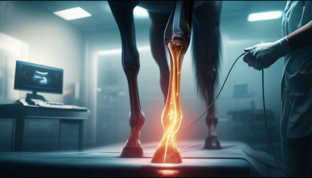
The tibial nerve, a major branch of the sciatic nerve, plays a vital role in the hindlimb's function. Blocking this nerve is often necessary to diagnose lameness affecting the lower leg and foot. An ultrasound-guided tibial nerve block involves using real-time ultrasound imaging to guide the placement of the needle and injection of local anesthetic near the tibial nerve. This ensures accurate and effective numbing of the target area.
- Preparation: The horse is gently restrained, and the injection site on the caudomedial aspect of the crus (lower leg) is clipped and cleaned.
- Imaging: A microconvex ultrasound probe is used to visualize the tibial nerve in a transverse section, about 8-10 cm proximal to the point of the hock. The nerve appears as an oval structure with distinct fascicles (bundles of nerve fibers).
- Needle Insertion: The needle is carefully inserted, guided by the ultrasound image, to the caudal aspect of the tibial nerve.
- Injection: Half of the anesthetic solution (typically 5-8 mL) is injected near the nerve. A second injection is made similarly, cranial to the probe, to ensure complete coverage of the nerve.
- Confirmation: The veterinarian confirms the correct placement and spread of the anesthetic solution around the tibial nerve using ultrasound.
The Future of Lameness Diagnosis: Precision and Accuracy
Ultrasound-guided nerve blocks represent a significant advancement in equine lameness diagnosis. By providing real-time visualization and precise needle placement, this technique improves the accuracy and effectiveness of nerve blocks while minimizing potential risks. This translates to faster, more reliable diagnoses, ultimately leading to better outcomes for horses experiencing lameness.