Light Therapy's Secret Weapon: How Imaging Reveals the Immune Boost
"See how scientists are using cutting-edge imaging to unlock the power of photodynamic therapy and its effect on your body's defenses."
Photodynamic therapy (PDT) is becoming increasingly recognized as a minimally invasive cancer treatment. In the United States, an earlier version of PDT using Porfimer sodium (Photofrin) gained FDA approval for treating conditions like Barrett's esophagus with high-grade dysplasia and certain types of obstructing cancers. However, early photosensitizers came with drawbacks, including prolonged skin photosensitivity and limited selectivity in targeting tumors, pushing researchers to explore more advanced alternatives.
One promising next-generation photosensitizer is 2-[1-hexyloxyethyl]-2 devinyl pyropheophorbide-a (HPPH). HPPH has shown excellent safety and effectiveness in treating various cancers while minimizing skin photosensitization. Scientists are particularly interested in how HPPH interacts with the body's immune system during PDT, but studying these interactions requires advanced techniques to observe the process in real-time and with minimal disturbance to the tissue.
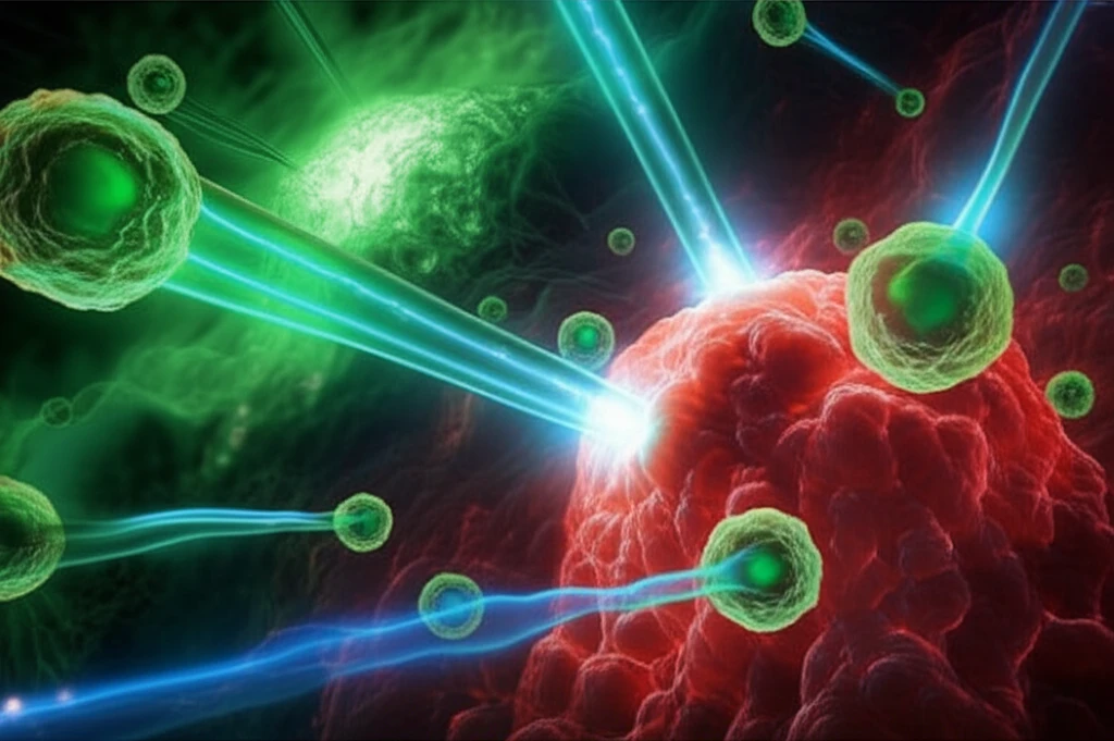
Now, researchers are employing innovative optical imaging strategies to noninvasively examine how photosensitizers distribute within tumors and how the body responds to HPPH-mediated PDT. By using these advanced imaging methods, scientists are gaining valuable insights into optimizing PDT for more effective cancer treatment and potentially unlocking new ways to harness the immune system's power.
The Eye in the Tumor: Watching HPPH at Work
Researchers used optical imaging to track HPPH's distribution and effects on EMT6 tumors in mice. They administered HPPH intravenously and used wide-field fluorescence imaging to observe that the drug accumulated more in the tumor than in surrounding healthy tissue. They observed approximately a 2-3 fold difference between tumor and surrounding normal tissue.
- Neutrophil Surge: A significant and rapid increase in Gr1+ cells was observed in response to the therapy. The accumulation peaked at 24 hours post-irradiation before decreasing at 48 hours.
- Vascular Damage: Reduced Gr1+ cell density at 48 hours strongly correlated with functional damage to the tumor's blood vessels, indicated by decreased perfusion.
- Neutrophil Activation: About 90% of the anti-Gr1+ cell population co-localized with anti-CD11b labeling, confirming that most of these cells were neutrophils.
- Antigen Presentation: A two-fold increase in MHC-II+ cells was observed 24 hours post-PDT. Further analysis revealed that a greater fraction of Gr1+ cells were expressing MHC-II, suggesting that HPPH-PDT stimulates neutrophils to develop an antigen-presenting phenotype.
A New Frontier in Cancer Treatment: Seeing is Believing
This research demonstrates the power of real-time imaging to understand the complex interactions between photodynamic therapy and the immune system. By visualizing these processes, scientists can optimize PDT protocols and potentially develop new strategies to enhance the body's natural defenses against cancer.
While techniques like flow cytometry offer advantages in identifying cell types, in vivo imaging provides a unique perspective by allowing researchers to study inflammatory responses in minimally perturbed tissue, preserving the natural architecture and cell interactions. This approach provides a measure of validation for the in vivo imaging strategy as an assay for host cell populations.
As imaging technologies continue to advance, we can expect even more detailed insights into the mechanisms of PDT and other cancer therapies, paving the way for more effective and personalized treatment strategies.