Knee Stability After Surgery: What You Need to Know About Recovery
"A comprehensive guide to understanding post-operative imaging, surgical procedures, and rehabilitation for patellofemoral instability."
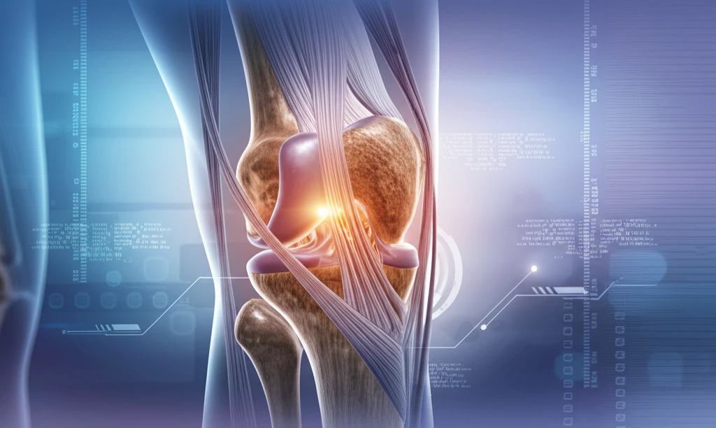
The knee is a complex joint. It relies on both bone and soft tissue to function correctly. When things go wrong with the patellofemoral joint (the area where your kneecap sits), it can lead to instability or persistent pain.
If you're dealing with patellofemoral issues, careful assessment through clinical exams and imaging is key. Cross-sectional imaging like MRI plays a vital role in planning any potential surgery.
Surgical treatments for patellofemoral problems can address soft tissue, bone issues, or both. This article explains what happens after surgery, focusing on imaging results of the knee's extensor mechanism. We'll cover common procedures, why they're done, and possible complications, empowering you to understand your recovery.
Understanding Post-operative Procedures and Imaging
After knee surgery, imaging (especially MRI) is essential to see how well the procedure worked and to spot any potential problems. The images will show the surgical site and any changes made to the surrounding tissues.
- Lateral Release and Medial Reefing: A lateral release involves cutting the tight tissues on the outer side of your kneecap. Medial reefing tightens the tissues on the inner side. Imaging may show subtle changes in the soft tissues.
- MPFL Reconstruction: The medial patellofemoral ligament (MPFL) is a crucial stabilizer of the kneecap. If it's damaged, reconstruction involves replacing it with a graft. Imaging will show the new ligament and the screws or anchors used to hold it in place.
- Distal Realignment Procedures: These surgeries address the alignment of the lower leg. The tibial tuberosity (the bony bump below your kneecap) is moved to improve the kneecap's tracking. Imaging shows the new position of the tuberosity and any hardware used.
- Trochleoplasty: This procedure reshapes the trochlear groove (the groove in your femur where the kneecap sits) to provide better stability. Imaging will reveal the altered shape of the groove.
Working Together for a Successful Recovery
Knee surgery is a significant step, but understanding what to expect afterward can make the recovery process smoother. By knowing the types of surgeries, what imaging reveals, and potential complications, you can be an active participant in your care. Partner with your orthopedic surgeon and physical therapist to create a personalized plan that gets you back to your active life with confidence. Your active involvement significantly contributes to a more effective and satisfying recovery journey.