Kidney Vision: Ultrasound Reveals Hidden Blood Flow
"New microscopy sharpens our view of tiny vessels, promising earlier disease detection."
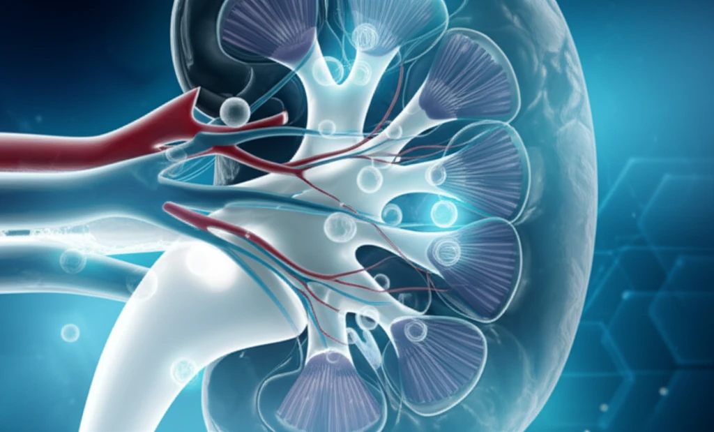
Imagine peering into the intricate network of blood vessels within your kidneys, spotting potential problems long before they cause serious damage. This level of detailed visualization is becoming a reality thanks to groundbreaking advancements in ultrasound technology. Traditionally, capturing images of the smallest vessels, known as microvasculature, has been a challenge.
A new study published in Scientific Reports details how researchers are pushing the boundaries of ultrasound localization microscopy (ULM) to image and assess the microvasculature in rat kidneys. This technique, which involves tracking individual microbubbles as they travel through the bloodstream, offers unprecedented spatial resolution, potentially revolutionizing the way we diagnose and monitor kidney diseases.
This article will break down the key findings of this study, explaining how ULM works, what challenges the researchers overcame, and why this technology holds so much promise for the future of kidney health. If you're interested in learning about the latest advancements in medical imaging and how they could impact early disease detection, keep reading.
How Does Ultrasound Localization Microscopy Work?
ULM is a technique that allows researchers to visualize blood vessels at a resolution far exceeding the limits of conventional ultrasound. It relies on injecting tiny gas-filled bubbles, called microbubbles, into the bloodstream. These microbubbles act as contrast agents, reflecting sound waves and making them visible on ultrasound images.
- Injection: Microbubbles are injected into the bloodstream.
- Imaging: An ultrasound system emits sound waves into the kidney.
- Detection: The microbubbles reflect these sound waves, creating echoes that are detected by the ultrasound system.
- Tracking: Sophisticated algorithms track the movement of individual microbubbles over time.
- Mapping: By mapping the locations of thousands of microbubbles, a detailed image of the microvasculature is created.
What's Next for Ultrasound Microscopy?
This study demonstrates the significant potential of ULM for visualizing and assessing the microvasculature in vivo. By overcoming challenges related to organ motion and developing advanced processing techniques, the researchers have paved the way for future applications of this technology in clinical settings.
While this research focused on rat kidneys, the principles of ULM can be applied to other organs as well. Future studies could explore the use of ULM for:
<ul><li>Detecting early signs of kidney damage in patients with diabetes or hypertension.</li><li>Monitoring the effectiveness of treatments for kidney disease.</li><li>Improving the diagnosis and treatment of tumors.</li><li>Visualizing blood flow in other organs, such as the heart and brain.</li></ul>