Futuristic kidney transplant ultrasound scan

Kidney Transplant Health: Can Ultrasound Predict Problems?

"New research explores how a special type of ultrasound, shear-wave sonoelastography, might help doctors spot problems early in kidney transplants."


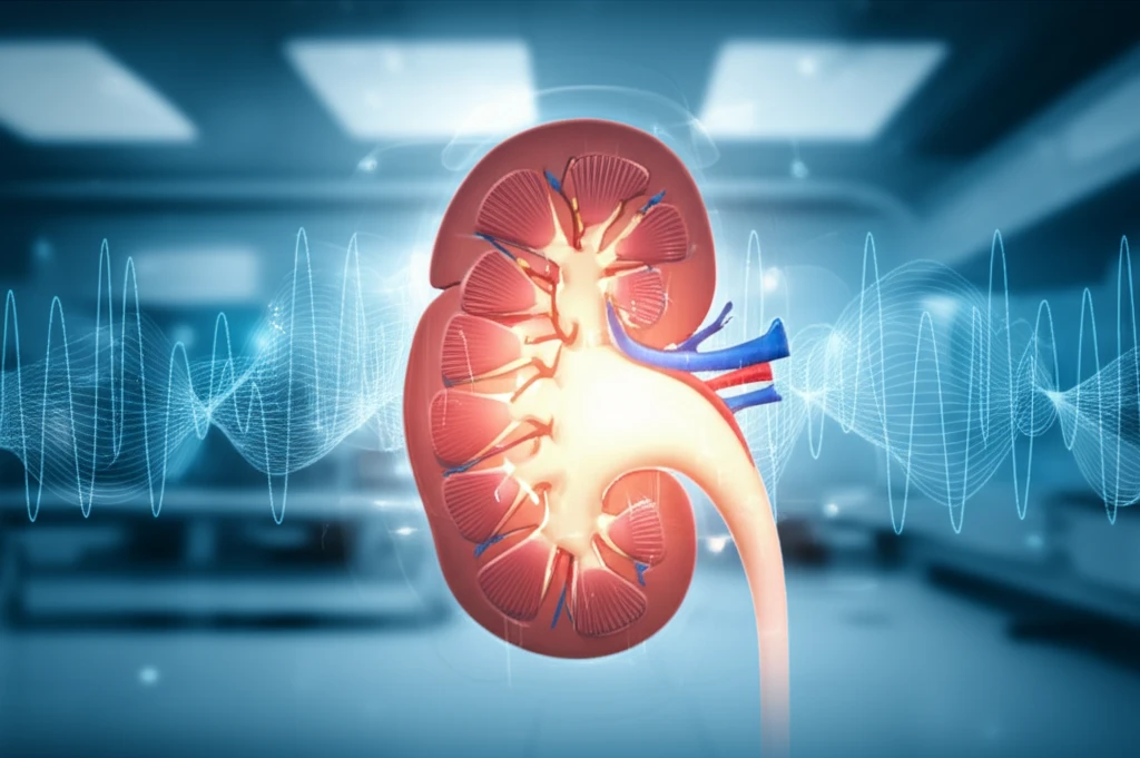
After a kidney transplant, keeping a close watch on the new organ is critical. Regular check-ups help ensure everything is working as it should. Traditionally, doctors use methods like standard ultrasounds, blood tests, and sometimes biopsies to assess the kidney's health. But imagine if there was a way to get even more information without the need for invasive procedures. That's where sonoelastography comes in.

Sonoelastography is an innovative imaging method that allows doctors to evaluate the stiffness of tissues. Approved by the FDA for liver diagnostics, this technique has demonstrated the ability to differentiate between healthy and cirrhotic liver tissue effectively. Beyond the liver, sonoelastography is being explored for its potential in diagnosing cancers and evaluating conditions like atherosclerosis.

A study published in "Radiology" in 2018, investigates whether shear-wave sonoelastography can distinguish between a stable renal allograft and acute or chronic allograft dysfunction, and correlate stiffness values with key indicators and biopsy results.

What is Shear-Wave Sonoelastography and How Does It Work?

Futuristic kidney transplant ultrasound scan

Shear-wave sonoelastography is a non-invasive ultrasound technique that assesses tissue stiffness. It works by sending shear waves into the tissue and measuring how quickly these waves travel. Stiffer tissues cause the waves to move faster, while softer tissues slow them down. This information is then translated into an image or numerical value, providing doctors with insights into the tissue's condition.

The "Radiology" study aimed to evaluate the effectiveness of shear-wave sonoelastography in identifying different types of kidney allograft dysfunction. The study involved 60 patients who had undergone kidney transplants. These patients were divided into three groups based on clinical assessments: those with stable allografts (functioning properly), those with acute allograft dysfunction (sudden issues), and those with chronic allograft dysfunction (long-term problems).

  • Stable Allograft: Kidneys functioning well for at least six months, with good eGFR (estimated glomerular filtration rate) and normal creatinine levels.
  • Acute Allograft Dysfunction: A sudden decline in kidney function, indicated by decreased eGFR, increased creatinine, and/or proteinuria (protein in the urine).
  • Chronic Allograft Dysfunction: A gradual worsening of kidney function over more than three months, also characterized by reduced eGFR, elevated creatinine, and proteinuria.
The study compared parenchymal stiffness (the stiffness of the kidney tissue) among the three groups. They also looked at correlations between stiffness measurements and other indicators like the resistive index (RI), serum creatinine levels, estimated glomerular filtration rate (eGFR), and findings from renal biopsies where available.

The Future of Kidney Transplant Monitoring

Shear-wave sonoelastography shows promise as a valuable tool in kidney transplant monitoring. By providing a non-invasive way to assess tissue stiffness, it could help doctors detect problems earlier, guide treatment decisions, and potentially reduce the need for invasive biopsies. Further research with larger patient groups and diverse populations will help solidify its role in improving long-term outcomes for kidney transplant recipients.

About this Article -

This article was crafted using a human-AI hybrid and collaborative approach. AI assisted our team with initial drafting, research insights, identifying key questions, and image generation. Our human editors guided topic selection, defined the angle, structured the content, ensured factual accuracy and relevance, refined the tone, and conducted thorough editing to deliver helpful, high-quality information.See our About page for more information.

Everything You Need To Know

1

What is shear-wave sonoelastography, and how does it help in evaluating kidney transplant health?

Shear-wave sonoelastography is a non-invasive ultrasound technique used to assess tissue stiffness. In the context of kidney transplants, it helps evaluate the health of the transplanted kidney (renal allograft) by measuring how quickly shear waves travel through the kidney tissue. Stiffer tissues indicate potential problems such as dysfunction, while softer tissues suggest healthier tissue. This method allows doctors to detect issues earlier, guiding treatment decisions and potentially reducing the need for invasive biopsies.

2

How does shear-wave sonoelastography differ from traditional methods used to monitor kidney transplant health?

Traditional methods for monitoring kidney transplant health include standard ultrasounds, blood tests (like creatinine and eGFR measurements), and sometimes biopsies. Shear-wave sonoelastography offers a significant advantage because it is non-invasive and provides additional information about the kidney tissue's stiffness. This can help detect subtle changes that might not be apparent with standard ultrasounds or blood tests alone, potentially allowing for earlier intervention and improved outcomes. Biopsies, which are invasive, are often used but carry inherent risks, making the non-invasive nature of shear-wave sonoelastography particularly valuable.

3

What were the key findings of the "Radiology" study regarding shear-wave sonoelastography and kidney transplants?

The "Radiology" study aimed to determine if shear-wave sonoelastography could differentiate between stable renal allografts and those experiencing acute or chronic allograft dysfunction. The study found correlations between the kidney tissue stiffness values obtained using shear-wave sonoelastography and key indicators of kidney function, such as eGFR, creatinine levels, and biopsy results. The research suggested that shear-wave sonoelastography could potentially be used to identify different types of kidney allograft dysfunction, helping doctors better manage and treat complications post-transplant.

4

Can you explain the difference between stable allograft, acute allograft dysfunction, and chronic allograft dysfunction in the context of a kidney transplant?

In a kidney transplant, a stable allograft refers to a transplanted kidney that is functioning well for at least six months, indicated by a good eGFR and normal creatinine levels. Acute allograft dysfunction involves a sudden decline in kidney function, marked by a decrease in eGFR, an increase in creatinine, and/or proteinuria. Chronic allograft dysfunction represents a gradual worsening of kidney function over more than three months, also characterized by reduced eGFR, elevated creatinine, and proteinuria. These classifications help doctors assess the severity and nature of any issues with the transplanted kidney, guiding appropriate treatment strategies.

5

What is the future outlook for shear-wave sonoelastography in the field of kidney transplant monitoring?

Shear-wave sonoelastography shows promise as a valuable tool in kidney transplant monitoring. By providing a non-invasive assessment of tissue stiffness, it could help doctors detect problems earlier, guide treatment decisions, and potentially reduce the need for invasive biopsies. Future research involving larger patient groups and diverse populations will be crucial to solidify its role. Further studies will help refine its use, optimize its parameters, and integrate it into the standard of care to improve long-term outcomes for kidney transplant recipients.

Newsletter Subscribe

Subscribe to get the latest articles and insights directly in your inbox.