Glowing kidney transplant surrounded by sound waves visualizing tissue health

Kidney Transplant Health: Can Shear-Wave Sonoelastography Help?

"A new study explores how shear-wave sonoelastography can improve the detection and management of kidney transplant dysfunction."


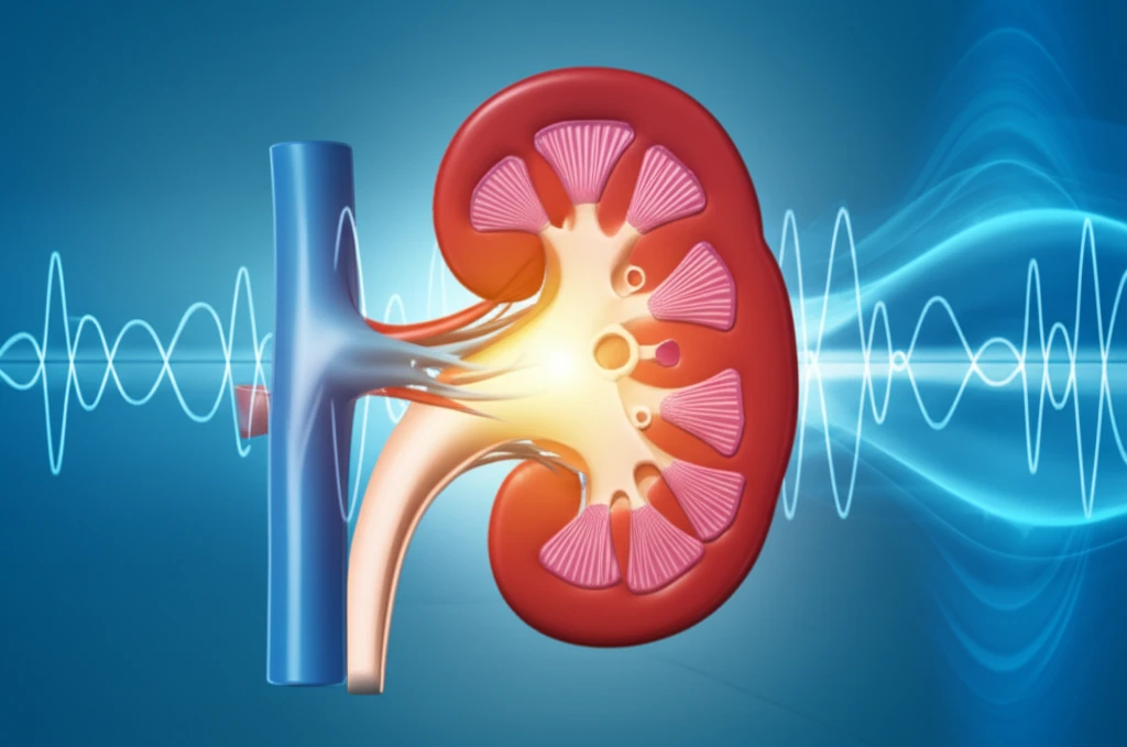
After a kidney transplant, ensuring the new organ functions well is crucial. Doctors use various methods to monitor the health of the transplanted kidney, known as an allograft. Traditionally, this involves blood tests, ultrasound imaging, and sometimes biopsies, which can be invasive. Researchers are always looking for ways to improve these evaluations.

Shear-wave sonoelastography is emerging as a promising non-invasive technique. It measures tissue stiffness, offering clues about the organ's condition. Think of it like feeling for firmness to detect problems, but with sound waves. This method has shown promise in diagnosing issues in other organs, and now, researchers are exploring its potential in kidney transplants.

A recent study published in "Radiology" investigated whether shear-wave sonoelastography could differentiate between healthy transplanted kidneys and those experiencing acute or chronic dysfunction. The study also examined how these measurements correlated with other standard health indicators, providing a comprehensive view of the allograft's health.

How Does Shear-Wave Sonoelastography Work in Kidney Transplants?

Glowing kidney transplant surrounded by sound waves visualizing tissue health

The study, conducted at Indraprastha Apollo Hospital in New Delhi, involved 60 patients who had undergone kidney transplants. These patients were categorized into three groups: those with stable allografts, those with acute allograft dysfunction, and those with chronic allograft dysfunction. The categorization was based on clinical assessments and laboratory results.

During the shear-wave sonoelastography procedure, a specialized ultrasound machine was used to measure the stiffness of the kidney tissue. Six measurements were taken in different areas of the transplanted kidney, and the average stiffness was calculated for each patient. These measurements were then compared across the three patient groups.

  • Stable Allograft: Kidneys functioning well with consistent eGFR (estimated glomerular filtration rate) above 50 mL/min over the previous six months.
  • Acute Allograft Dysfunction: Sudden decline in kidney function, indicated by reduced eGFR, increased serum creatinine (by at least 0.3 mg/dL or 50%), and presence of proteinuria.
  • Chronic Allograft Dysfunction: Gradual worsening of kidney function over more than three months, with eGFR below 50 mL/min, elevated serum creatinine, and proteinuria.
Researchers also collected data on other relevant health indicators, including resistive index (RI), serum creatinine levels, and eGFR. They then analyzed how these indicators correlated with the stiffness measurements obtained through shear-wave sonoelastography. In a subset of patients, biopsy findings were also considered to provide a direct comparison with the sonoelastography results.

The Future of Kidney Transplant Monitoring

This study provides valuable insights into the potential of shear-wave sonoelastography as a tool for monitoring kidney transplant health. While further research is needed, these findings suggest that this non-invasive technique could play an important role in the early detection and management of allograft dysfunction, ultimately improving outcomes for transplant recipients.

About this Article -

This article was crafted using a human-AI hybrid and collaborative approach. AI assisted our team with initial drafting, research insights, identifying key questions, and image generation. Our human editors guided topic selection, defined the angle, structured the content, ensured factual accuracy and relevance, refined the tone, and conducted thorough editing to deliver helpful, high-quality information.See our About page for more information.

Everything You Need To Know

1

What is shear-wave sonoelastography and why is it being explored for kidney transplants?

Shear-wave sonoelastography is a non-invasive technique that measures tissue stiffness using sound waves. It's being explored for kidney transplants because it can potentially help in assessing the health of the transplanted kidney, or allograft, without the need for invasive procedures like biopsies. By measuring the stiffness of the kidney tissue, doctors can gain insights into whether the allograft is functioning properly or experiencing acute or chronic dysfunction. This method is similar to palpation, but instead of using touch, it uses sound waves to assess tissue properties.

2

How does shear-wave sonoelastography differentiate between a stable allograft and one with dysfunction?

Shear-wave sonoelastography measures the stiffness of the kidney tissue. A stable allograft, which is a kidney functioning well, typically exhibits a certain range of stiffness values. Acute allograft dysfunction, characterized by a sudden decline in kidney function, and chronic allograft dysfunction, which involves a gradual worsening over time, may present with altered stiffness measurements compared to a stable allograft. The study mentioned categorizes patients based on clinical assessments, laboratory results, and stiffness measurements obtained through shear-wave sonoelastography. Analyzing these stiffness differences, in conjunction with indicators like eGFR and serum creatinine, allows for differentiation.

3

What are the key health indicators, besides tissue stiffness, that are considered alongside shear-wave sonoelastography in kidney transplant monitoring?

Besides tissue stiffness measured by shear-wave sonoelastography, key health indicators include the estimated glomerular filtration rate (eGFR), serum creatinine levels, and the resistive index (RI). eGFR indicates how well the kidneys are filtering waste, while serum creatinine is a measure of kidney function. The resistive index, often measured via Doppler ultrasound, reflects blood flow resistance within the kidney. These indicators, when correlated with shear-wave sonoelastography results, provide a comprehensive assessment of the allograft's health, aiding in the detection and management of potential dysfunction.

4

What are the implications of using shear-wave sonoelastography for early detection of allograft dysfunction?

Early detection of allograft dysfunction through shear-wave sonoelastography can lead to timely interventions, potentially improving outcomes for kidney transplant recipients. If acute allograft dysfunction or chronic allograft dysfunction is detected early, clinicians can adjust immunosuppressant medications, manage complications, and prevent further damage to the allograft. This can help prolong the life of the transplanted kidney and reduce the need for re-transplantation or dialysis. By providing a non-invasive method for regular monitoring, shear-wave sonoelastography could significantly enhance the long-term management of kidney transplant patients. However, to fully confirm this benefit prospective clinical trials are needed.

5

What is the significance of eGFR, serum creatinine, and proteinuria in the context of kidney transplant health, and how do these relate to allograft dysfunction?

eGFR (estimated glomerular filtration rate), serum creatinine, and proteinuria are crucial indicators of kidney function and play a significant role in identifying allograft dysfunction. eGFR measures how well the kidneys are filtering waste from the blood; a decline suggests impaired function. Serum creatinine is a waste product that healthy kidneys remove; elevated levels indicate that the kidneys aren't working efficiently. Proteinuria, the presence of protein in the urine, signifies damage to the kidney's filtering units. In acute allograft dysfunction, eGFR decreases, serum creatinine increases significantly, and proteinuria may be present. Chronic allograft dysfunction is characterized by a gradual decline in eGFR, elevated serum creatinine, and persistent proteinuria. These markers, when assessed together, provide a comprehensive picture of allograft health, helping clinicians distinguish between stable allografts and those experiencing dysfunction.

Newsletter Subscribe

Subscribe to get the latest articles and insights directly in your inbox.