Kidney Health Unveiled: How Cutting-Edge Imaging is Revolutionizing Renal Transplant Care
"New research highlights how advanced imaging techniques are transforming the way we monitor and manage kidney transplant patients, offering hope for improved outcomes and a better quality of life."
Kidney transplantation is a life-altering procedure, offering a new lease on life for individuals battling end-stage renal disease. However, the journey doesn't end with the transplant. Ensuring the long-term health and functionality of the transplanted kidney, or allograft, is a continuous process. This requires diligent monitoring and timely intervention to address any complications, such as allograft dysfunction.
Allograft dysfunction can manifest in various forms, ranging from acute rejection to chronic damage. Accurate and early detection is crucial to prevent graft failure and optimize patient outcomes. Traditionally, monitoring relies on blood tests and imaging techniques. However, recent advancements in imaging technology are providing a more comprehensive and detailed assessment of allograft health.
This article delves into the groundbreaking use of shear-wave sonoelastography, a non-invasive imaging technique, in evaluating renal allograft dysfunction. We will explore how this technology is changing the way healthcare professionals monitor and manage kidney transplant patients, offering a new level of precision in diagnosing and managing allograft dysfunction. We will also explore the study's implications for the future of renal care.
Unveiling the Power of Shear-Wave Sonoelastography in Renal Transplant Care
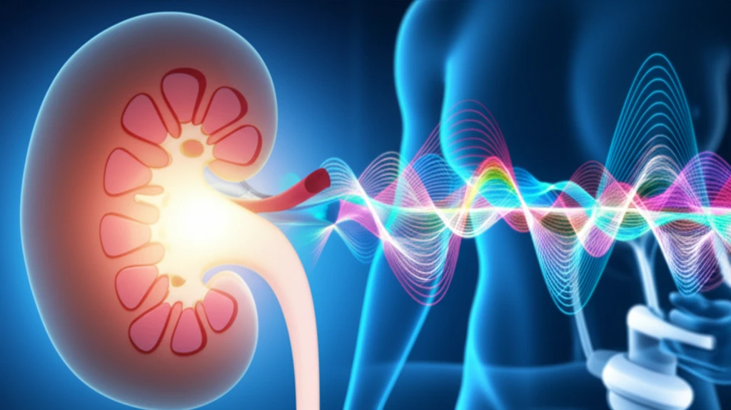
Shear-wave sonoelastography is an innovative ultrasound-based technique that assesses the stiffness of tissues. This is particularly useful in evaluating the renal allograft, as changes in tissue stiffness can indicate underlying issues such as fibrosis or inflammation. Unlike traditional ultrasound, which primarily provides anatomical information, shear-wave sonoelastography offers a functional assessment of the allograft, providing valuable insights into its health.
- Stable Allografts: Showed lower stiffness values, indicating healthy tissue.
- Acute Allograft Dysfunction: Exhibited moderately increased stiffness, often associated with inflammation or early rejection.
- Chronic Allograft Dysfunction: Displayed significantly higher stiffness values, reflecting the presence of fibrosis and chronic damage.
The Future of Renal Transplant Care
The findings of this study underscore the potential of shear-wave sonoelastography to transform the landscape of renal transplant care. By providing a non-invasive, accurate, and comprehensive assessment of allograft health, this technology empowers healthcare professionals to make more informed decisions, leading to improved patient outcomes and a better quality of life. As research continues to evolve, shear-wave sonoelastography promises to be a cornerstone in the future of renal transplant care.