Is Your 'Never Warm' Port-Wine Stain Actually Something Else?
"A new case study suggests that the traditional diagnostic feature of Port-Wine Stains might not be as reliable as we thought, especially when heat is involved."
Port-wine stains (PWSs) are commonly known as congenital vascular malformations, characterized by dilated capillaries in the skin. These typically appear as pink or purple marks at birth, often on the face or neck, and are notably considered 'never warm' to the touch. But what happens when a PWS isn't congenital, and—more surprisingly—is warm?
A recent case study sheds light on acquired PWS, a rare condition that mirrors the appearance and microscopic features of congenital PWS but develops later in life. This study highlights a unique instance where an acquired PWS presented with local heat, challenging the traditional diagnostic criteria.
This article delves into the specifics of this intriguing case, exploring the methods used to diagnose it and discussing the implications for how we identify and understand PWS. It also addresses the questions of what to do if the temperature reading differs and how it could refine diagnostic process.
The Curious Case of the Warm Port-Wine Stain
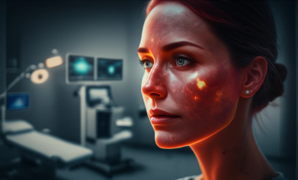
A 46-year-old woman presented with slowly enlarging reddish-purple patches on her left cheek and neck that had developed over the course of a year. Unlike typical PWS, these lesions felt mildly warm and slightly swollen during examination. The woman had been previously misdiagnosed with rosacea. She confirmed that she had no history of such marks, trauma, or family history of PWS, and took no medications.
- Skin Biopsy: Microscopic examination revealed dilated blood vessels in the dermis, consistent with PWS.
- Laser Doppler Perfusion Imaging (LDPI): This non-invasive imaging technique mapped blood flow, revealing increased blood flow in the affected area, even in regions without visible symptoms.
- Laser Doppler Perfusion Measurement (LDPM): This test quantified skin temperature and microcirculatory blood perfusion, confirming elevated temperature and blood flow on the left side of her face and neck compared to the right.
- Immunohistochemical Staining: Testing ruled out arteriovenous malformation (AVM), another vascular condition that can cause similar symptoms.
Rethinking PWS Diagnosis: What Does This Mean for You?
This case not only highlights the rarity of acquired PWS but also challenges a long-standing diagnostic assumption. The presence of local heat in this case suggests that PWS can manifest differently than traditionally described.
For individuals with vascular lesions, it’s crucial to seek comprehensive evaluation that includes advanced imaging techniques like LDPI and LDPM. These tools can provide detailed information about blood flow and temperature, aiding in accurate diagnosis and treatment planning.
While pulsed dye laser therapy was considered for this patient, she opted to defer treatment. This decision underscores the importance of personalized care and informed decision-making in managing PWS and other vascular conditions. Further research and longer follow-up studies are needed to fully understand the implications of these findings and refine diagnostic approaches.