Brain with highlighted regions affected by insomnia

Is Your Brain on High Alert? How Insomnia Changes Brain Structure

"New research reveals how primary insomnia leads to gray matter hypertrophy, potentially explaining the persistent hyper-arousal associated with the sleep disorder."


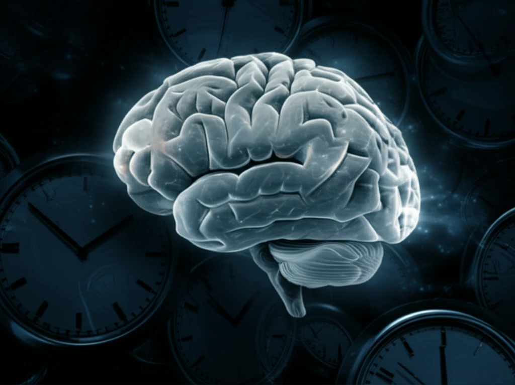
For many, insomnia is a frustratingly familiar struggle. The tossing and turning, the racing thoughts, the desperate clock-watching – it’s a nightly battle for millions. But what if insomnia is more than just a sleep disorder? What if it leaves a tangible mark on the very structure of your brain?

While past studies have hinted at brain abnormalities in individuals with primary insomnia (PI), these were often limited by small sample sizes and a focus on specific brain regions. Now, new research employing advanced surface-based morphometry (SBM) techniques offers a more comprehensive picture of how insomnia reshapes the brain.

This article will explore the key findings of this study, diving into how primary insomnia can lead to gray matter hypertrophy – an enlargement of brain tissue – in specific areas associated with crucial functions like emotional regulation, attention, and sensory processing. Understanding these structural changes could unlock new avenues for diagnosing and treating this pervasive sleep disorder.

Insomnia's Impact: Hyper-arousal and Brain Changes

Brain with highlighted regions affected by insomnia

The study, conducted by researchers at Chengdu University of Traditional Chinese Medicine and Southeast University, involved 67 patients with PI and 55 healthy controls. Using magnetic resonance imaging (MRI), the team meticulously examined the cortical morphology – the thickness and volume of the brain’s outer layer – in both groups.

The results revealed a striking difference: individuals with PI exhibited cortical thickening in several key regions, including:

  • Left orbital frontal cortex (OFC): Involved in emotional regulation and decision-making.
  • Right rostral anterior cingulate cortex (rACC): Plays a role in emotional processing and attention.
  • Left middle cingulate cortex (MCC): Contributes to attention control and emotional regulation.
  • Bilateral insula: Important for salience and emotional processing.
  • Left superior parietal lobule (SPL): Involved in spatial cognition and attention.
  • Right fusiform area (FFA): Associated with facial processing and visual recognition.
Furthermore, the study found increased cortical volume in the left OFC, right rACC, bilateral rostral middle frontal gyrus, and right FFA in PI patients. What’s particularly interesting is that cortical thickness in the right OFC and FFA positively correlated with the severity of insomnia symptoms, suggesting a relationship where greater thickening is related to more severe insomnia.

The Hyper-arousal Model: A New Perspective on Insomnia

These findings align with the hyper-arousal model of insomnia, which posits that individuals with PI experience a heightened state of alertness that interferes with their ability to fall and stay asleep. The observed gray matter hypertrophy in regions associated with attention, emotional regulation, and sensory processing may reflect the brain's attempt to compensate for this chronic state of hyper-arousal.

This study marks a significant step forward in understanding the neuropathology of primary insomnia. By identifying specific structural changes in the brain, researchers are paving the way for more targeted diagnostic and therapeutic interventions.

While further research is needed to fully elucidate the mechanisms underlying these brain changes and their long-term consequences, these findings offer hope for developing more effective treatments to alleviate the burden of insomnia and improve the lives of millions.

About this Article -

This article was crafted using a human-AI hybrid and collaborative approach. AI assisted our team with initial drafting, research insights, identifying key questions, and image generation. Our human editors guided topic selection, defined the angle, structured the content, ensured factual accuracy and relevance, refined the tone, and conducted thorough editing to deliver helpful, high-quality information.See our About page for more information.

This article is based on research published under:

DOI-LINK: 10.1007/s11682-018-9992-z, Alternate LINK

Title: Gray Matter Hypertrophy In Primary Insomnia: A Surface-Based Morphometric Study

Subject: Behavioral Neuroscience

Journal: Brain Imaging and Behavior

Publisher: Springer Science and Business Media LLC

Authors: Siyi Yu, Fen Feng, Qi Zhang, Zhifu Shen, Zhengyan Wang, Youping Hu, Liang Gong

Published: 2018-12-04

Everything You Need To Know

1

What is primary insomnia?

Primary insomnia (PI) is a sleep disorder characterized by difficulty falling asleep, staying asleep, or both, despite having adequate opportunities for sleep. Unlike insomnia caused by other medical conditions or substance use, primary insomnia isn't directly linked to another health problem. The study focuses on individuals diagnosed with PI, examining the structural changes in their brains compared to healthy controls.

2

What specific brain changes were identified in those with primary insomnia?

The study found that individuals with primary insomnia (PI) exhibited gray matter hypertrophy, or an enlargement of brain tissue, in several key regions of the brain. These regions include the Left orbital frontal cortex (OFC), Right rostral anterior cingulate cortex (rACC), Left middle cingulate cortex (MCC), Bilateral insula, Left superior parietal lobule (SPL), and Right fusiform area (FFA). Moreover, increased cortical volume was observed in the left OFC, right rACC, bilateral rostral middle frontal gyrus, and right FFA. These areas are associated with crucial functions like emotional regulation, attention, and sensory processing.

3

Why is gray matter hypertrophy important in relation to insomnia?

Gray matter hypertrophy is significant in the context of primary insomnia (PI) because it offers a new understanding of the disorder beyond just a sleep problem. The enlargement of brain tissue in areas related to emotional regulation, attention, and sensory processing may reflect the brain's adaptation to a chronic state of hyper-arousal, a key characteristic of PI. The correlation between greater cortical thickness in some regions and the severity of insomnia symptoms suggests a direct link between these structural changes and the experience of insomnia, opening new possibilities for diagnosis and treatment.

4

What is the hyper-arousal model of insomnia and how does this research connect to it?

The hyper-arousal model suggests that individuals with primary insomnia (PI) experience a heightened state of alertness that interferes with their ability to fall and stay asleep. The study's findings support this model by showing gray matter hypertrophy in brain regions associated with attention, emotional regulation, and sensory processing. This could be the brain’s response to the constant state of alertness, potentially creating a feedback loop where the brain works harder in these areas, leading to structural changes. This perspective shifts the focus from simply treating sleep difficulties to addressing the underlying brain changes associated with the hyper-arousal state.

5

What are the implications of these findings for managing and treating insomnia?

The implications of the study's findings are significant for the diagnosis and treatment of primary insomnia (PI). Understanding that PI involves structural changes in the brain opens up possibilities for more targeted interventions. For example, if the left orbital frontal cortex (OFC) shows changes, therapies could focus on emotional regulation. These changes may also lead to improved diagnostic tools using MRI to identify these brain structural changes and potentially to monitor the effectiveness of treatments. This research underscores the need for a more comprehensive approach to managing insomnia that considers both the sleep disturbance and the underlying neurological factors.

Newsletter Subscribe

Subscribe to get the latest articles and insights directly in your inbox.