Is Your Bone Density Test Telling the Whole Story? Unpacking Cortical Porosity
"Beyond Standard Scans: A Closer Look at How Micro-CT Scans Can Improve Bone Health Assessments for Osteoporosis and Fracture Risk"
Bone health is critical, but its complexity often gets reduced to a single number from a bone density test. While these tests are valuable for diagnosing osteoporosis and assessing fracture risk, they primarily focus on bone mineral density (BMD). However, the architecture of bone plays a vital role in its strength and resilience. One key aspect of bone architecture is cortical porosity, which refers to the tiny pores within the dense outer layer of bone. When these pores become too large or numerous, they can significantly weaken the bone and increase fracture risk, even if BMD appears normal.
Traditional bone density tests, such as DXA (dual-energy X-ray absorptiometry), have limitations in assessing cortical porosity. They provide a two-dimensional snapshot of bone density but can't fully capture the three-dimensional structure and porosity. This means that individuals with seemingly healthy BMD scores might still be at risk for fractures due to compromised bone architecture. As a result, researchers are exploring advanced imaging techniques to evaluate cortical porosity more accurately and comprehensively.
This article explores the importance of assessing cortical porosity in addition to bone mineral density for a more complete picture of bone health. We'll delve into advanced imaging methods, specifically high-resolution peripheral quantitative computed tomography (HR-pQCT) and synchrotron radiation micro-CT (SR-µCT), and how they can provide valuable insights for personalized osteoporosis management and fracture prevention.
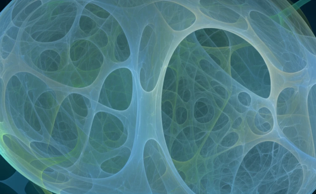
What is Cortical Porosity and Why Does It Matter?
Imagine a strong, sturdy building. Its strength depends not only on the amount of material used (like BMD) but also on how that material is arranged. Cortical bone makes up the outer shell of most bones, providing a protective layer and contributing significantly to overall bone strength. Cortical porosity refers to the presence of small holes or pores within this dense outer layer. A certain degree of porosity is normal and allows for nutrient exchange and bone remodeling. However, when porosity increases excessively, it compromises the bone's structural integrity, making it more susceptible to fractures.
- Fragility Indicator: Elevated cortical porosity is a red flag indicating increased bone fragility, independent of BMD.
- Fracture Risk: Higher porosity directly correlates with a greater risk of fractures, especially in the elderly.
- Loss and Age: Cortical porosity tends to increase with age, making older adults more vulnerable.
- Quantifiable Loss: Cortical porosity (Ct.Po) measures can reliably mark bone loss and fragility.
- Advanced Imaging: Modern imaging helps detect Ct.Po non-invasively, enhancing diagnostics.
The Future of Bone Health Assessment
Assessing cortical porosity holds immense promise for improving bone health assessment and fracture prevention. By incorporating advanced imaging techniques like HR-pQCT and SR-µCT into clinical practice, healthcare professionals can gain a more comprehensive understanding of bone strength and identify individuals at risk for fractures earlier. This personalized approach to osteoporosis management can lead to more effective interventions, such as lifestyle modifications, targeted exercises, and medications, to protect bone health and reduce the burden of fractures.