Is Oxygen the Key? Unlocking the Mysteries of Retinal Ischemia
"A deeper look at how oxygen levels affect vision in retinal vein occlusion and other ischemic conditions."
Recent studies using en face optical coherence tomography (OCT) have highlighted the variable nature of macular middle-retinal infarction (MMRI) in eyes experiencing retinal vein or arterial occlusion. This variability reflects differing levels of circulatory failure during the evolution of an infarct, challenging existing models.
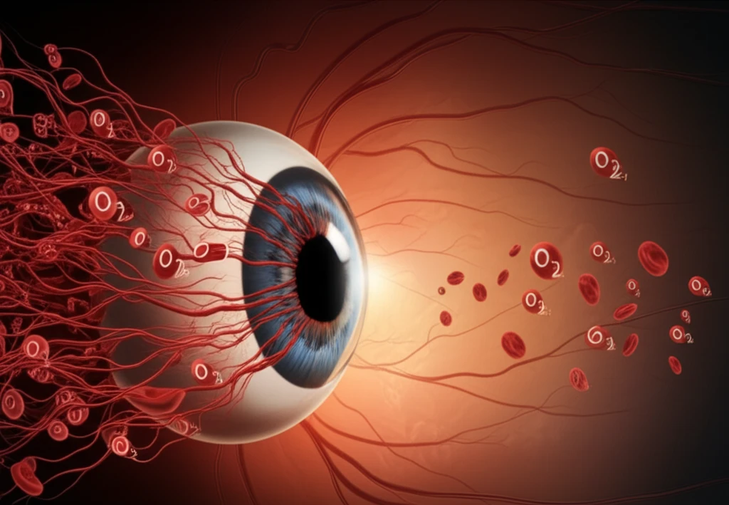
Researchers have proposed that MMRI stems from "misery perfusion," where the inner retina suffers from disproportionate oxygen extraction, leading to ischemic hypoxia and anoxia. This challenges the traditional understanding of tissue oxygenation, suggesting that the anatomic structure of macular arterioles and venules plays a critical role.
This article delves into the concept of oxygenation-based hypoperfusion maculopathy (OHM) to provide a broader perspective on ischemic dysfunction in the retina, moving beyond the limitations of terms like "perivenular PAMM" and addressing the role of oxygen levels in different retinal layers.
Beyond PAMM: Understanding Oxygenation-Based Hypoperfusion Maculopathy (OHM)
The term PAMM (“paracentral acute middle maculopathy”) has been used to describe specific lesions in CRVO and CRAO. However, this term may be limiting because OCT cannot directly identify hypoxic tissue. This means it might underestimate the full impact of inner retinal hypoperfusion. Furthermore, the expression "perivenular PAMM" is inherently contradictory.
- OHM Grade 1 (OHM1): Corresponds to the "fern-like" perivenular opacification seen in CRVO. The SIR is typically normoxic, while MMRI may show asymmetry.
- OHM Grade 2 (OHM2): Similar to "partial" CRAO, the SIR becomes predominantly hypoxic, and the MR is diffusely affected. Cotton-wool spots may appear.
- OHM Grade 3 (OHM3): Represents "complete" CRAO, where both the MR and SIR are anoxic, although some areas around third-order arterioles might be spared due to residual blood flow.
The Future of Retinal Ischemia Research
Moving forward, a deeper understanding of oxygen dynamics within the retina is crucial for developing targeted treatments for ischemic conditions. By recognizing the limitations of previous classifications and embracing the OHM framework, researchers and clinicians can better address the complexities of retinal vascular diseases.
Further research is needed to refine the OHM classification and explore potential therapeutic interventions that can improve oxygen delivery and reduce ischemic damage in the retina. This includes investigating strategies to enhance blood flow, reduce inflammation, and protect retinal cells from oxidative stress.
Ultimately, a comprehensive approach that considers the multifaceted nature of retinal ischemia, including oxygenation levels, vascular structure, and cellular responses, will lead to more effective treatments and improved outcomes for patients at risk of vision loss.