Cardiac MRI showing heart scar tissue

ICD Therapy: Can Cardiac Scars Predict If You Need One?

"New research uses cardiac MRI to analyze scar tissue, potentially improving how we determine who benefits most from ICDs."


Sudden cardiac death (SCD) due to ventricular arrhythmias is a major concern for individuals with ischemic cardiomyopathy (ICM) and nonischemic cardiomyopathy (NICM). An implantable cardioverter-defibrillator (ICD) is often prescribed as a preventative measure, delivering life-saving shocks to restore a normal heart rhythm.

However, ICDs aren't without risks and only a fraction of those who receive them ever actually need their shock therapy. This highlights the critical need for better risk stratification methods to accurately identify individuals who will truly benefit from an ICD, avoiding unnecessary procedures and potential complications for those who won't.

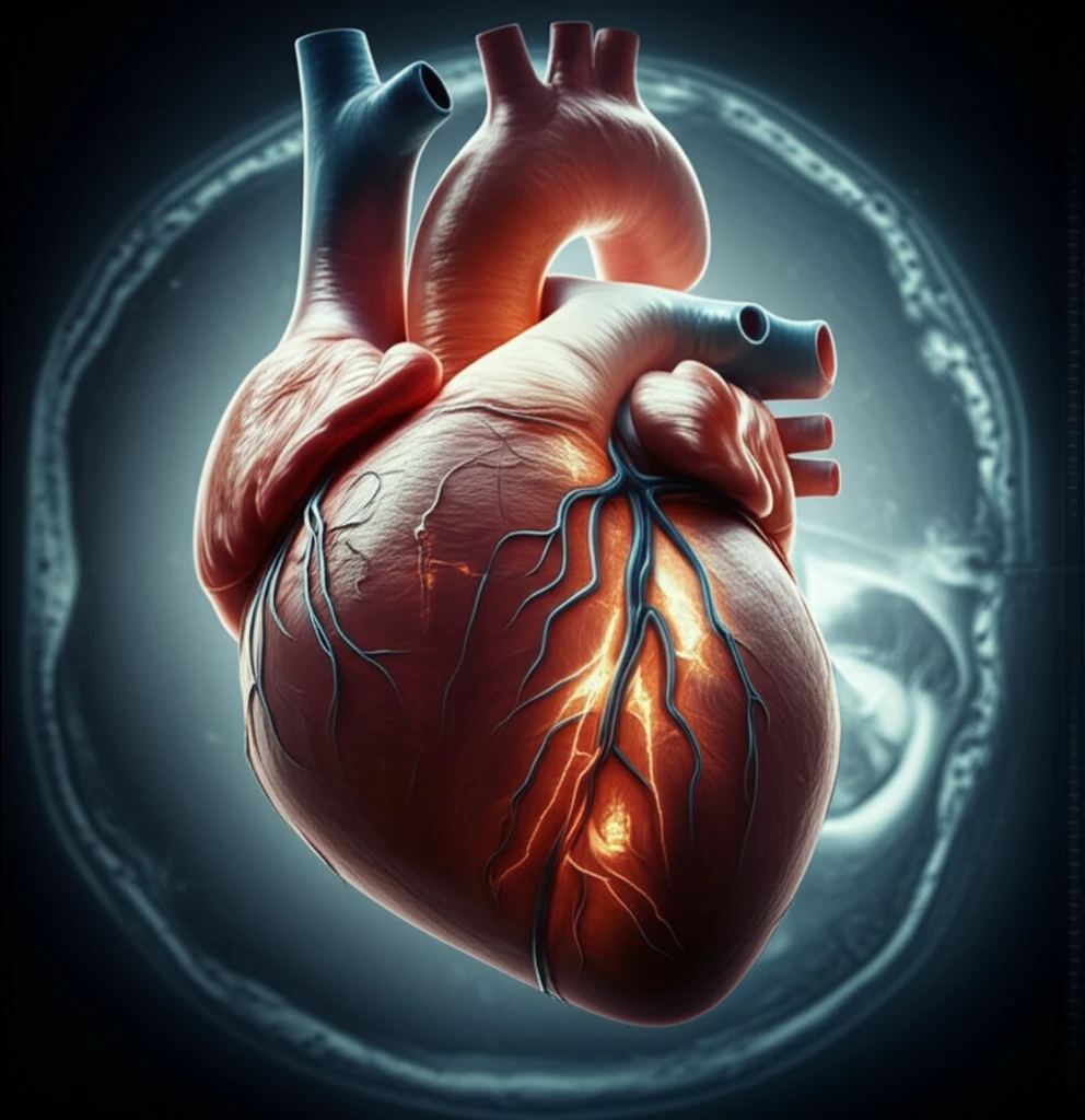
Enter cardiac magnetic resonance imaging (MRI) with late gadolinium enhancement (LGE). This advanced imaging technique allows doctors to visualize scar tissue within the heart muscle. The size, location, and characteristics of these scars can provide valuable insights into a person's risk of developing dangerous arrhythmias. This article explores recent research into how LGE border zones, the areas surrounding dense scar tissue, can be used to predict the success of ICD therapy, potentially refining patient selection and improving outcomes.

Cardiac Scars: A New Way to Predict ICD Success?

Cardiac MRI showing heart scar tissue

Researchers have been investigating the potential of LGE border zones as independent predictors of ventricular arrhythmias. While the presence and extent of LGE have been linked to predicting arrhythmic events, there's no universal agreement on the best method to analyze these border zones. To address this, a recent study compared four different algorithms for assessing LGE border zones and their ability to predict appropriate ICD therapy in ICM and NICM patients.

The study retrospectively included ICM and NICM patients who underwent LGE cardiac MRI before receiving an ICD. The researchers then used the four algorithms below to quantify the LGE border zones:

  • Expectation Maximization, Weighted Intensity, and A Priori Information (EWA)
  • Weighted Border Zone algorithm (WBZ)
  • Modified Full-Width Half-Maximum (mFWHM)
  • A 2-3SD threshold-based algorithm (2-3SD)
The algorithms were compared based on their ability to predict appropriate ICD therapy, with hazard ratios calculated per 1% increase in LGE. The findings revealed that in ICM patients with primary prophylactic ICDs, the EWA, WBZ, and mFWHM algorithms all successfully predicted ICD therapy. These algorithms also demonstrated a high negative predictive value of 92%, suggesting that patients with small LGE border zones may not benefit from an ICD.

What This Means for You

This research suggests that LGE border zone quantification can improve patient selection for primary prevention ICDs, especially in those with ICM. By accurately identifying individuals at low risk of arrhythmias, doctors can potentially avoid unnecessary ICD implantations and their associated complications.

While these findings are promising, it's important to note that the study also highlights the impact of the algorithm used for LGE border zone analysis. The predictive value of LGE border zone differed depending on the algorithm, emphasizing the need for standardized methods and further research to optimize LGE assessment.

If you're at risk for sudden cardiac death or have been diagnosed with ICM or NICM, discuss these findings with your cardiologist. Cardiac MRI with LGE could be a valuable tool in determining whether an ICD is the right choice for you. This information, combined with other clinical factors, can help you and your doctor make informed decisions about your heart health and treatment options.

About this Article -

This article was crafted using a human-AI hybrid and collaborative approach. AI assisted our team with initial drafting, research insights, identifying key questions, and image generation. Our human editors guided topic selection, defined the angle, structured the content, ensured factual accuracy and relevance, refined the tone, and conducted thorough editing to deliver helpful, high-quality information.See our About page for more information.

This article is based on research published under:

DOI-LINK: 10.1161/circimaging.116.006105, Alternate LINK

Title: Cardiovascular Magnetic Resonance To Predict Appropriate Implantable Cardioverter Defibrillator Therapy In Ischemic And Nonischemic Cardiomyopathy Patients Using Late Gadolinium Enhancement Border Zone

Subject: Cardiology and Cardiovascular Medicine

Journal: Circulation: Cardiovascular Imaging

Publisher: Ovid Technologies (Wolters Kluwer Health)

Authors: Robert Jablonowski, Uzma Chaudhry, Jesper Van Der Pals, Henrik Engblom, Håkan Arheden, Einar Heiberg, Katherine C. Wu, Rasmus Borgquist, Marcus Carlsson

Published: 2017-09-01

Everything You Need To Know

1

What is an Implantable Cardioverter-Defibrillator (ICD) and why is it important?

An Implantable Cardioverter-Defibrillator (ICD) is a device implanted in the chest to monitor the heart rhythm. If a life-threatening arrhythmia occurs, the ICD delivers an electrical shock to restore a normal heart rhythm, preventing sudden cardiac death. It is a preventative measure primarily for individuals with conditions like ischemic cardiomyopathy (ICM) and nonischemic cardiomyopathy (NICM). The importance lies in its life-saving potential, but the device isn't without risks, and not everyone who receives one will require its therapy. This highlights the need for improved risk stratification to identify those who will truly benefit.

2

What is Cardiac MRI with late gadolinium enhancement (LGE) and why is it relevant?

Cardiac magnetic resonance imaging (MRI) with late gadolinium enhancement (LGE) is an advanced imaging technique that allows doctors to visualize scar tissue within the heart muscle. It's a key tool for evaluating the risk of ventricular arrhythmias. This technique is significant because the characteristics of the cardiac scars, including their size, location, and the border zones around them, can provide valuable insights into a person's risk. It enables doctors to assess the extent of damage to the heart tissue which is an important factor in determining whether someone would benefit from an ICD.

3

What are LGE border zones and why are they significant?

LGE border zones are the areas surrounding dense scar tissue within the heart muscle, as visualized by cardiac MRI with late gadolinium enhancement. These zones are important because recent research has focused on using the characteristics of these areas to predict the success of Implantable Cardioverter-Defibrillator therapy. The algorithms such as Expectation Maximization, Weighted Intensity, and A Priori Information (EWA), Weighted Border Zone algorithm (WBZ), and Modified Full-Width Half-Maximum (mFWHM) can identify patients who are at low risk of arrhythmias. This could avoid unnecessary ICD implantations and their associated complications.

4

What are Ischemic Cardiomyopathy (ICM) and Nonischemic Cardiomyopathy (NICM), and why are they mentioned in this context?

Ischemic cardiomyopathy (ICM) is a condition where the heart muscle is weakened due to reduced blood flow, often caused by coronary artery disease. Nonischemic cardiomyopathy (NICM) refers to heart muscle diseases not caused by reduced blood flow. Both conditions can lead to an increased risk of ventricular arrhythmias and sudden cardiac death (SCD). Because of this high risk, individuals with ICM or NICM are often considered for an Implantable Cardioverter-Defibrillator (ICD). Identifying the correct patients for the therapy is critical to prevent both unnecessary procedures and to ensure that those who need the device receive it.

5

What are the different algorithms used to analyze the LGE border zones, and what role do they play?

The algorithms mentioned – Expectation Maximization, Weighted Intensity, and A Priori Information (EWA), Weighted Border Zone algorithm (WBZ), Modified Full-Width Half-Maximum (mFWHM), and a 2-3SD threshold-based algorithm (2-3SD) – are methods used to quantify the LGE border zones. These algorithms analyze the data from cardiac MRI with late gadolinium enhancement to determine the extent and characteristics of scar tissue. The study compared these algorithms to identify the best approach for predicting the success of Implantable Cardioverter-Defibrillator (ICD) therapy in patients with ischemic cardiomyopathy (ICM) and nonischemic cardiomyopathy (NICM). They were compared based on their ability to predict the need for appropriate ICD therapy. The success of these algorithms can help avoid unnecessary ICD implants, reducing patient risk and improving outcomes.

Newsletter Subscribe

Subscribe to get the latest articles and insights directly in your inbox.