Healing Hearts: How Fibroblast Activation Impacts Recovery After a Heart Attack
"Uncover the critical role of fibroblast activation in cardiac remodeling and fibrosis post-myocardial infarction, and how targeted treatments could improve patient outcomes."
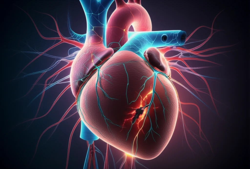
A heart attack, or myocardial infarction (MI), triggers a cascade of events that can lead to long-term heart damage. While the immediate focus is on restoring blood flow, the subsequent healing process involves significant changes in the heart's structure and function. Key players in this remodeling are fibroblasts (Fb), cells responsible for maintaining the heart's connective tissue. After an MI, these fibroblasts become activated, contributing to the formation of scar tissue and the development of fibrosis, which can impair the heart's ability to pump efficiently.
Fibrosis, the excessive accumulation of connective tissue, is a double-edged sword. In the short term, it helps to stabilize the damaged area. However, over time, fibrosis can stiffen the heart muscle, leading to heart failure. Understanding how fibroblasts differentiate and contribute to fibrosis in different regions of the heart after an MI is crucial for developing targeted therapies to promote healing and prevent adverse outcomes.
Recent research has shed light on the regional differences in fibroblast activation and fibrosis following an MI. A study published in Scientific Reports investigated these processes in a pig model, revealing that while fibroblast activation occurs throughout the left ventricle, the extent of fibrosis varies depending on the location relative to the scar tissue. This article delves into these findings, exploring the implications for future treatments aimed at optimizing heart recovery after an MI.
Fibroblast Activation: A Whole-Heart Phenomenon
The study highlighted that fibroblast activation, a process where fibroblasts transform into myofibroblasts (cells with contractile properties), occurs throughout the left ventricle after an MI. This activation is largely driven by transforming growth factor-beta 1 (TGF-β1), a protein that stimulates fibroblast differentiation and is released in response to injury and increased mechanical stress on the heart wall. This suggests that the entire left ventricle undergoes a degree of remodeling as a consequence of the heart attack.
- Scar Tissue: Primarily composed of collagen I and III, forming highly cross-linked fibers.
- Adjacent Myocardium (Mladjacent): Significant interstitial fibrosis with increased collagen I, and perivascular fibrosis.
- Remote Myocardium (MIremote): Minimal fibrosis, similar to healthy tissue.
Targeting Fibrosis: A Path to Better Heart Health
The study's findings underscore the importance of understanding the regional differences in fibroblast activation and fibrosis after an MI. While broad strategies to reduce TGF-β1 signaling may help to dampen overall fibroblast activation, targeted therapies that address local factors driving fibrosis in the Mladjacent region may be more effective in preventing adverse remodeling. Future research should focus on identifying these local cues and developing strategies to modulate fibroblast behavior in a region-specific manner, ultimately leading to improved outcomes for patients recovering from heart attacks.