
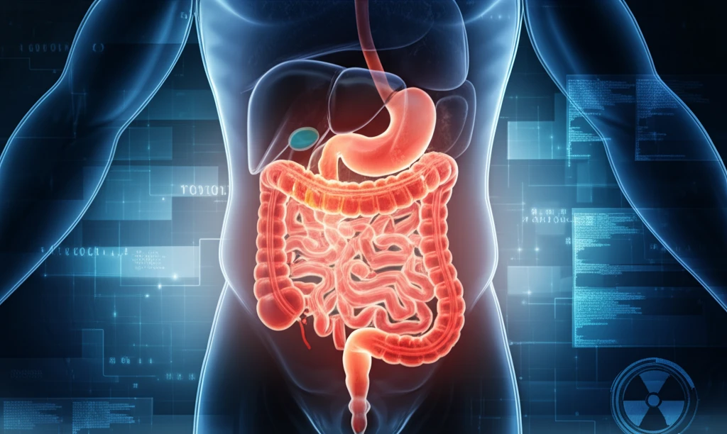
Gut Check: Unlocking the Secrets of Your Digestive System Through Nuclear Medicine

"From swallowing studies to liver function, discover how nuclear medicine provides crucial insights into gastrointestinal health and helps doctors diagnose and treat complex digestive disorders."
Ever wonder how doctors get a detailed look inside your digestive system without invasive procedures? The answer lies in nuclear medicine, a specialized field that uses small amounts of radioactive materials to diagnose and treat a variety of diseases. In the realm of gastrointestinal (GI) health, nuclear medicine offers unique insights that traditional imaging techniques often can't provide.
Nuclear medicine procedures are like having tiny spies that travel through your body, emitting signals that can be detected by special cameras. These signals help doctors visualize the function of different organs and identify abnormalities that might be causing you discomfort or illness. Think of it as a high-tech way to see what's really going on beneath the surface.
This article will explore the various applications of nuclear medicine in gastroenterology. We'll delve into the specific tests used to evaluate different parts of your digestive system, from your esophagus to your liver, and explain how these tests can help your doctor pinpoint the source of your problems and develop an effective treatment plan.
Digestive Health by the Numbers
Gastrointestinal disorders represent a substantial share of global healthcare burden, affecting hundreds of millions of people worldwide. Digestive conditions range from common motility issues to life-threatening gastrointestinal bleeding and rare cancers such as gastrointestinal stromal tumors. While precise prevalence figures vary by region and condition, GI diseases consistently rank among the leading causes of physician visits and hospitalizations. Nuclear medicine has emerged as an important diagnostic tool for evaluating many of these conditions, offering functional imaging that complements structural studies.
How Nuclear Medicine Works for GI Imaging
Nuclear medicine therapies can typically be performed as outpatient procedures, with few side effects and radiation exposure kept within safe limits for the general public. Radionuclide imaging is standard practice for detecting acute gastrointestinal bleeding, among other applications. However, researchers have identified numerous pitfalls and limitations across GI nuclear medicine, including challenges in hepatobiliary and gastrointestinal system imaging that can affect diagnostic accuracy. These limitations underscore the need for careful technique and awareness of confounding factors during interpretation.
The Roots of GI Nuclear Medicine
Gastrointestinal nuclear medicine has been formalized as a distinct subspecialty, with foundational knowledge compiled in texts such as Radiology Fundamentals: Introduction to Imaging & Technology. Gastrointestinal scintigraphy has long been recognized as an integral and important component of assessing gastrointestinal function, including motor disorders. Pharmacologic interventions during scintigraphy have also been developed to aid in diagnosis and treatment, expanding the diagnostic toolkit available to clinicians studying GI motility and transit.
A Deep Dive into Nuclear Medicine Techniques for GI Health
Nuclear medicine offers a range of diagnostic tools tailored to different parts of the digestive system. Here's a closer look at some of the key procedures:
- Early detection of salivary gland dysfunction.
- Non-invasive assessment of gland function.
- Helps differentiate between different salivary gland disorders.
New Frontiers in GI Nuclear Imaging
Researchers continue to refine nuclear medicine techniques for localizing gastrointestinal bleeding, with multiple approaches developed to assess intermittent bleeding or identify the unknown site of bleeding. A 2024 study highlighted that commonly used medications, including cannabis products and weight loss drugs, can impact the accuracy of gastric emptying scans, urging physicians to be more attuned to these side effects. Additionally, diagnostic applications in nuclear medicine are expanding for gastrointestinal stromal tumors, which, although rare, represent the most common mesenchymal gastrointestinal neoplasm and involve complex epidemiologic and genetic factors.
Challenges and Practical Limitations
While nuclear medicine GI scans are widely used, practical challenges persist in clinical implementation. Patient preparation is critical: scans require fasting for at least four hours, with overnight fasting preferred, and medications that delay gastric emptying must be stopped well in advance. The specificity of findings can be limited, and clinicians must carefully consider the clinical context when interpreting results to avoid misdiagnosis.
Progress and Competing Approaches
Gastrointestinal nuclear medicine has made significant progress in recent years, driven by advances in instrumentation and the development of new radiopharmaceuticals. For conditions like gastrointestinal stromal tumors, the landscape has evolved beyond nuclear imaging alone, with medical oncology playing an increasingly central role in treatment and tumor response assessment. Imatinib and subsequent targeted therapies have transformed GIST management, creating a multidisciplinary approach in which nuclear medicine is one component among several diagnostic and therapeutic modalities.
Empowering Your Digestive Health Journey
Nuclear medicine has revolutionized the way we understand and manage gastrointestinal disorders. By providing detailed functional information, these techniques enable doctors to make more accurate diagnoses and develop personalized treatment plans. If you're experiencing digestive issues, talk to your doctor about whether nuclear medicine might be right for you. Remember, knowledge is power, and understanding your digestive system is the first step towards a healthier, happier you.
A Multidisciplinary View of GI Nuclear Medicine
Leading cancer centers now employ multidisciplinary teams for gastrointestinal conditions, integrating surgeons, medical oncologists, nuclear medicine specialists, endocrinologists, interventional radiologists, and pathologists into cohesive care units. Gastrointestinal nuclear medicine encompasses a broad range of studies, including hepatobiliary scintigraphy, gastric emptying assessment, hepatic and splenic imaging, and bowel hot spot detection. This breadth reflects the versatility of nuclear medicine as a diagnostic framework, though it also demands substantial expertise across multiple organ systems and disease processes.
Where GI Nuclear Medicine Is Headed
An editorial in the nuclear medicine literature has posed the pointed question of whether gastrointestinal nuclear medicine is truly making progress, prompting reflection on the field's trajectory and future direction. Continued development of new radiopharmaceuticals and imaging technologies offers promise for improved diagnostic accuracy and patient outcomes. However, realizing this potential will require sustained investment in research and training to ensure that advances translate into meaningful clinical benefits.
Systemic Hurdles Facing the Field
Gastrointestinal nuclear medicine operates within a broader healthcare landscape shaped by competing diagnostic modalities, including advanced CT, MRI, and endoscopic techniques. Access to nuclear medicine services remains uneven across healthcare systems, with availability often concentrated in larger medical centers. Balancing the unique functional insights offered by nuclear imaging against the structural detail of alternative modalities remains an ongoing challenge for clinicians and healthcare administrators alike.
Real-World Protocols and Patient Experience
Nuclear medicine offers a number of studies to diagnose diseases of the hepatobiliary system and broader gastrointestinal tract, grounded in decades of clinical application. In practice, patients undergoing GI nuclear medicine scans follow specific protocols, such as a solid study requiring a minimum four-hour fast and the oral administration of a radiopharmaceutical like Tc 99m Sulfur Colloid. These structured protocols help ensure diagnostic reliability while reflecting the practical realities of nuclear medicine imaging in real-world clinical settings.