Surreal illustration of granular cell tumor on a child's back.

Granular Cell Tumors in Children: A Rare but Important Diagnosis

"Understanding childhood granular cell tumors: A case report and comprehensive overview for early detection and treatment."


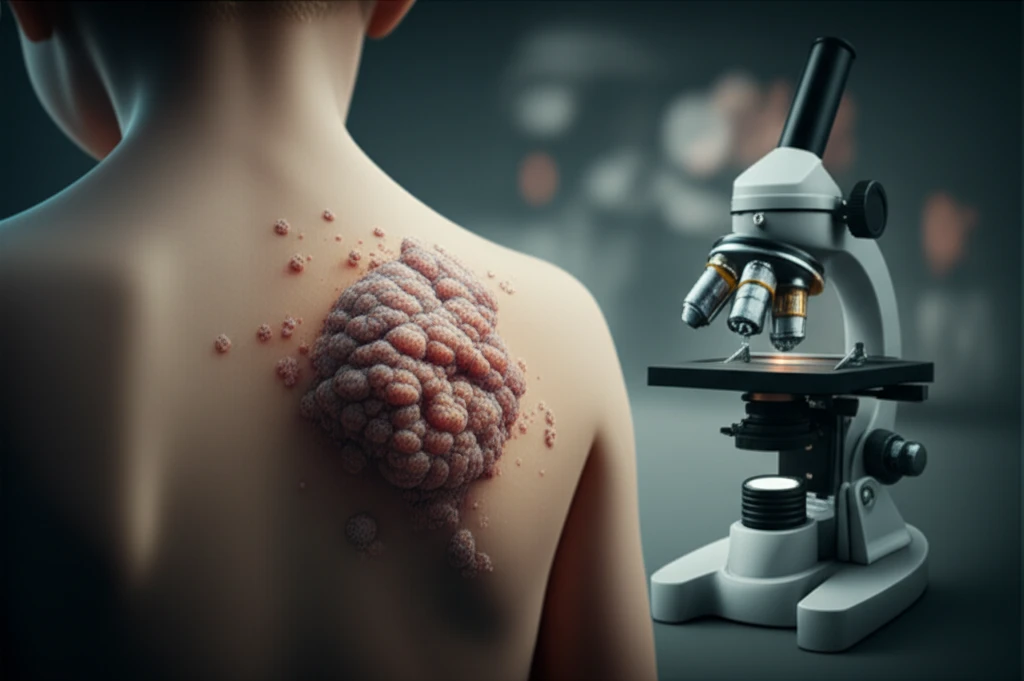
Granular cell tumors (GCTs) are benign neoplasms characterized by the presence of cells with distinctive granular eosinophilic cytoplasm when viewed under a microscope. While generally benign, their rarity in children makes accurate diagnosis and appropriate management crucial. These tumors pose a diagnostic challenge due to their varied presentation and the need to differentiate them from other, more common lesions.

Originally, GCTs were thought to arise from muscle cells, leading to the now-outdated term "myoblastoma." Today, it is widely accepted that GCTs originate from Schwann cells, which are responsible for producing the myelin sheath that insulates nerve fibers. Despite this understanding, the exact cause and development of GCTs remain areas of ongoing research.

This article delves into a recent case of a childhood GCT and provides a comprehensive overview of the condition, highlighting key diagnostic features, treatment strategies, and the importance of awareness among healthcare professionals. By increasing understanding of this rare entity, we hope to facilitate earlier detection and improved outcomes for affected children.

What are the Key Features of Granular Cell Tumors in Children?

Surreal illustration of granular cell tumor on a child's back.

A recent case highlights the typical presentation and diagnostic pathway for GCTs in children. An 11-year-old boy presented with an asymptomatic nodule on his back that had been growing progressively over six months. Physical examination revealed a mobile, non-tender, oval nodule measuring 1.5 cm in diameter.

The diagnostic process involves a combination of clinical evaluation and pathological examination. Key steps include:

  • Clinical Examination: Careful assessment of the nodule's size, location, consistency, and associated symptoms.
  • Histological Study: Microscopic examination of a tissue sample obtained through biopsy. GCTs are characterized by large cells with abundant granular eosinophilic cytoplasm and centrally located nuclei.
  • Immunohistochemistry: Specialized staining techniques to identify specific proteins within the tumor cells. GCTs typically show positivity for S100 protein, a marker associated with Schwann cell differentiation.
In the reported case, histological examination of the excised nodule confirmed the diagnosis of GCT, with characteristic cellular morphology and S100 positivity. Conservative surgical excision was performed, and the patient experienced a full recovery. This case underscores the importance of considering GCT in the differential diagnosis of solitary skin nodules in children.

Why is Early Detection of Granular Cell Tumors Crucial?

While GCTs are generally benign, early detection and appropriate management are essential for several reasons. Accurate diagnosis helps to avoid unnecessary anxiety and aggressive treatments. Surgical excision, when indicated, can be performed in a conservative manner, minimizing the risk of complications and preserving function. Furthermore, awareness of the rare possibility of malignant transformation is crucial for long-term monitoring.

About this Article -

This article was crafted using a human-AI hybrid and collaborative approach. AI assisted our team with initial drafting, research insights, identifying key questions, and image generation. Our human editors guided topic selection, defined the angle, structured the content, ensured factual accuracy and relevance, refined the tone, and conducted thorough editing to deliver helpful, high-quality information.See our About page for more information.

Everything You Need To Know

1

What exactly are Granular Cell Tumors (GCTs), and where do they come from?

Granular Cell Tumors (GCTs) are benign neoplasms characterized by cells with a distinct granular, eosinophilic cytoplasm, as observed under a microscope. Initially, they were mistakenly believed to originate from muscle cells, hence the old term "myoblastoma." Current understanding points to Schwann cells as the origin, these cells produce the myelin sheath which insulates nerve fibers. Although the exact development process of GCTs is still under investigation.

2

How are Granular Cell Tumors (GCTs) diagnosed in children?

The diagnosis of GCTs in children involves a multi-step process. It begins with a clinical examination where the healthcare provider assesses the nodule's size, location, consistency, and any associated symptoms. Next is a histological study, which involves microscopic examination of a tissue sample obtained through a biopsy. The presence of large cells with abundant granular eosinophilic cytoplasm and centrally located nuclei is a key indicator. Immunohistochemistry is then used with specialized staining techniques to identify specific proteins within the tumor cells. GCTs typically show positivity for S100 protein, a marker associated with Schwann cell differentiation.

3

What are the key steps in diagnosing a Granular Cell Tumor (GCT)?

The diagnostic process involves a combination of clinical evaluation and pathological examination. The key steps include Clinical Examination which involves assessing the nodule's size, location, consistency, and associated symptoms. A Histological Study which is the microscopic examination of a tissue sample obtained through biopsy. Lastly, Immunohistochemistry uses specialized staining techniques to identify specific proteins within the tumor cells. GCTs typically show positivity for S100 protein, a marker associated with Schwann cell differentiation.

4

Why is it so important to detect Granular Cell Tumors (GCTs) early in children?

Early detection of Granular Cell Tumors (GCTs) is crucial because, although they are generally benign, early diagnosis helps in avoiding unnecessary anxiety and potentially aggressive treatments. Early detection also enables conservative surgical excision which helps minimize the risk of complications. Furthermore, awareness of the rare possibility of malignant transformation is crucial for long-term monitoring.

5

Can you describe a real-life case of a child diagnosed with a Granular Cell Tumor (GCT)?

In a recent case, an 11-year-old boy was diagnosed with a GCT. The boy presented with an asymptomatic nodule on his back that had been progressively growing for six months. Physical examination revealed a mobile, non-tender, oval nodule measuring 1.5 cm in diameter. After clinical evaluation, a histological study was performed and confirmed the diagnosis of GCT, with characteristic cellular morphology and S100 positivity. Conservative surgical excision was performed, and the patient fully recovered. This case emphasizes the importance of considering GCT in the differential diagnosis of solitary skin nodules in children.

Newsletter Subscribe

Subscribe to get the latest articles and insights directly in your inbox.