ADC mapping reveals hidden tumor cells in the brain.

Glioblastoma's Stealth Spread: How New MRI Techniques Spot Hidden Tumor Growth

"Discover how diffusion MRI and ADC mapping are changing the game in glioblastoma treatment by revealing tumor infiltration that standard scans miss."


Glioblastoma, an aggressive form of brain cancer, presents a significant challenge in treatment due to its ability to spread into surrounding brain tissue. Traditional MRI scans, which rely on contrast enhancement to visualize tumors, can be misleading, especially when anti-VEGF (vascular endothelial growth factor) therapies are used. These therapies reduce blood vessel permeability, making the tumor appear smaller on scans, even if it continues to infiltrate the brain.

A groundbreaking study published in Neuro-Oncology explored the use of diffusion MRI, a technique that measures water molecule movement in tissues, to detect this 'stealth' spread of glioblastoma after treatment with cediranib, an anti-VEGF drug. The research highlights the potential of apparent diffusion coefficient (ADC) mapping to reveal areas of tumor infiltration that are not visible on standard contrast-enhanced MRI.

This article delves into the findings of this study, explaining how ADC mapping works, what it reveals about glioblastoma's behavior under anti-VEGF treatment, and why this new approach could be crucial for improving treatment strategies and patient outcomes. We'll break down the complex science into understandable terms, revealing how this innovative imaging technique offers a more accurate picture of tumor activity.

ADC Mapping: Unmasking Hidden Tumor Activity

ADC mapping reveals hidden tumor cells in the brain.

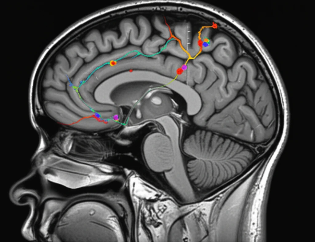
Diffusion MRI measures the movement of water molecules within brain tissue. In areas of high cell density, like tumors, water movement is restricted. ADC mapping quantifies this restriction, assigning lower ADC values to areas where water diffusion is limited, indicating a higher concentration of tumor cells.

Researchers in the Neuro-Oncology study used ADC mapping to analyze MRI scans of 30 glioblastoma patients undergoing treatment with cediranib. They established a patient-specific threshold to differentiate between normal brain tissue and areas with abnormally low ADC values, suggesting tumor infiltration.

The key findings of the study revealed:
  • Increased Infiltration: The percentage of the FLAIR hyperintensity (an area of abnormal signal on MRI) comprised of low ADC values increased significantly over time during cediranib treatment. This suggests that while the contrast-enhanced portion of the tumor might appear to shrink, the tumor was actually spreading into surrounding tissue.
  • Hidden Growth: Visualization of low ADC voxels revealed regions of tumor growth that were not visible on contrast-enhanced MRI. This confirmed the hypothesis that anti-VEGF therapies can mask tumor infiltration.
  • Potential for Prediction: Changes in ADC values were associated with progression-free survival, suggesting that ADC mapping could be used to predict treatment response.
In essence, ADC mapping acts like a 'tumor cell density detector,' highlighting areas where cancer cells are actively infiltrating, even when standard MRI scans provide a misleadingly optimistic picture.

The Future of Glioblastoma Treatment Monitoring

The Neuro-Oncology study provides compelling evidence that ADC mapping can offer a more accurate assessment of glioblastoma behavior during anti-VEGF therapy. By revealing hidden tumor infiltration, this technique could help clinicians make more informed treatment decisions.

However, the researchers emphasize the need for further studies to validate these findings and refine the use of ADC mapping in clinical practice. Future research should focus on establishing standardized ADC thresholds and correlating ADC changes with histological confirmation of tumor infiltration.

Ultimately, the integration of diffusion MRI and ADC mapping into routine glioblastoma monitoring could lead to earlier detection of tumor progression, more personalized treatment strategies, and improved outcomes for patients battling this challenging disease. This approach exemplifies the shift towards more precise and informative imaging techniques in cancer care.

About this Article -

This article was crafted using a human-AI hybrid and collaborative approach. AI assisted our team with initial drafting, research insights, identifying key questions, and image generation. Our human editors guided topic selection, defined the angle, structured the content, ensured factual accuracy and relevance, refined the tone, and conducted thorough editing to deliver helpful, high-quality information.See our About page for more information.

Everything You Need To Know

1

What is Glioblastoma, and why is it a challenge to treat?

Glioblastoma is an aggressive form of brain cancer known for its ability to infiltrate surrounding brain tissue. This makes treatment challenging because the tumor's spread can be difficult to detect using standard imaging techniques. The article highlights this challenge and how new imaging methods are improving detection.

2

What is ADC mapping, and how does it work?

ADC mapping is a specialized type of diffusion MRI. It measures the movement of water molecules within brain tissue. Tumors, due to their high cell density, restrict water movement. ADC mapping quantifies this restriction, with lower ADC values indicating areas of higher tumor cell concentration. The article explains how ADC mapping can reveal tumor infiltration that is not visible on standard MRI scans, especially when anti-VEGF therapies are used.

3

How do anti-VEGF therapies affect the appearance of glioblastoma on MRI scans?

Anti-VEGF therapies, such as cediranib, are designed to reduce blood vessel permeability, potentially making tumors appear smaller on standard MRI scans. However, this doesn't mean the tumor isn't growing; it can still be infiltrating the brain tissue. The use of anti-VEGF therapies can mask the extent of glioblastoma infiltration, making it challenging to assess treatment effectiveness using traditional MRI methods. The study's findings emphasize the need for more accurate imaging techniques to monitor tumor behavior during this treatment.

4

What were the key findings of the study using ADC mapping?

The study revealed increased infiltration of the tumor, as indicated by a rise in low ADC values during cediranib treatment. This finding suggests that while the contrast-enhanced part of the tumor might seem to shrink, the tumor continues to spread. Moreover, ADC mapping revealed regions of tumor growth that were not visible on conventional MRI scans, validating the hypothesis that anti-VEGF therapies can conceal tumor infiltration. Also, alterations in ADC values have been linked to progression-free survival, showing that ADC mapping may have the potential to predict the outcome of treatments.

5

What are the potential implications of using ADC mapping in glioblastoma treatment?

The implications of ADC mapping are significant. By providing a more detailed picture of glioblastoma infiltration, clinicians can make better-informed treatment decisions. The potential to predict treatment response and monitor tumor behavior more accurately could lead to earlier interventions and improved patient outcomes. The technique offers a more reliable way to assess the effectiveness of treatments like anti-VEGF therapies, addressing the limitations of standard MRI scans.

Newsletter Subscribe

Subscribe to get the latest articles and insights directly in your inbox.