Brain with glowing tumor cells illuminated by fluorescent light, superimposed on MRI scans

Glioblastoma Breakthrough: Shining a Light on Tumor Cells Beyond MRI Detection

"New research reveals how a fluorescent dye can highlight hidden tumor cells in brain cancer, even when MRI scans appear clear."


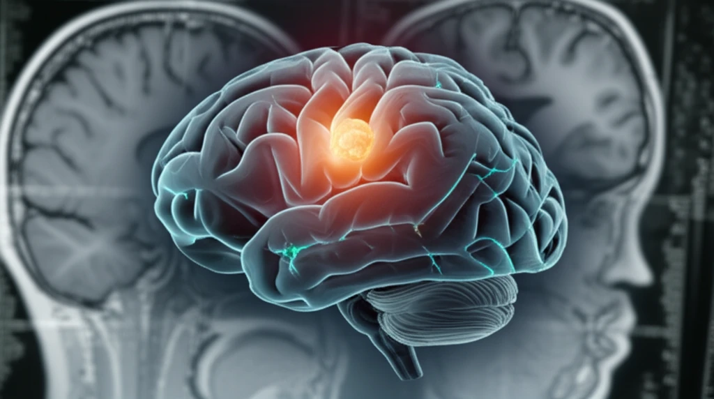
Glioblastoma multiforme (GBM) is one of the most aggressive forms of brain cancer. Treating it often involves surgery to remove as much of the tumor as possible. Surgeons use various tools and techniques to guide them, including magnetic resonance imaging (MRI). MRI helps visualize the tumor, but sometimes, cancer cells can hide in plain sight, evading detection by conventional imaging.

A fluorescent substance called 5-aminolevulinic acid (5-ALA) is emerging as a valuable tool in GBM surgery. When administered to patients, 5-ALA causes tumor cells to glow under a special blue light, making them easier to see and remove. This technique has significantly improved the extent of tumor resection, leading to better outcomes for patients.

However, a perplexing phenomenon has been observed during 5-ALA-guided surgery: fluorescence in the walls of the brain's ventricles (fluid-filled spaces) even when MRI scans show no sign of tumor involvement. A new study published in Oncology Reports investigates this unexpected fluorescence, shedding light on the presence of tumor cells beyond the reach of MRI detection.

Unveiling Hidden Tumor Cells with 5-ALA Fluorescence

Brain with glowing tumor cells illuminated by fluorescent light, superimposed on MRI scans

The study, led by researchers at Yonsei University College of Medicine in South Korea, involved 19 patients with newly diagnosed GBM located near the lateral ventricle. During surgery, the surgeons carefully examined the ventricular walls for fluorescence after administering 5-ALA. They collected tissue samples from areas with and without fluorescence, even if the MRI scans showed no tumor presence.

The researchers found that 11 out of the 19 patients (57.9%) exhibited 5-ALA-induced fluorescence in the ventricular wall. What was striking was that in some of these areas, standard MRI scans showed no signs of tumor. This raised a critical question: What was causing the fluorescence in the absence of visible tumor?

  • Intraoperative Observation: Surgeons used microscopes with special filters to identify areas of fluorescence in the ventricle walls during GBM surgery.
  • Tissue Sampling: 25 ventricular wall tissue samples were collected, regardless of whether they showed fluorescence or had visible tumor involvement on MRI.
  • Histopathological Analysis: These samples underwent detailed examination to determine if tumor cells were present, and advanced techniques like immunohistochemistry and genetic analysis were used.
The histopathological analysis revealed that of the 11 samples with 5-ALA-induced fluorescence, 5 (45.5%) contained tumor cells. More surprisingly, the remaining 6 (54.5%) were free of tumor cells. Conversely, all 14 samples that lacked 5-ALA-induced fluorescence showed no evidence of tumor cells. These findings suggest that 5-ALA fluorescence can detect glioma cells in the ventricle wall even when MRI images appear clear.

Implications and Future Directions for Glioblastoma Treatment

This study highlights the potential of 5-ALA fluorescence to reveal hidden tumor cells that MRI might miss. This could lead to more complete tumor removal during surgery, potentially improving patient outcomes. However, the study also raises questions about what causes fluorescence in the absence of tumor cells. Further research is needed to understand the nature of this unexpected ventricular wall fluorescence and to optimize the use of 5-ALA in GBM surgery. More studies including molecular characterization, of these falsely fluorescing samples are warranted.

About this Article -

This article was crafted using a human-AI hybrid and collaborative approach. AI assisted our team with initial drafting, research insights, identifying key questions, and image generation. Our human editors guided topic selection, defined the angle, structured the content, ensured factual accuracy and relevance, refined the tone, and conducted thorough editing to deliver helpful, high-quality information.See our About page for more information.

Everything You Need To Know

1

What is Glioblastoma multiforme, and why is it considered such a serious condition?

Glioblastoma multiforme (GBM) is a highly aggressive form of brain cancer. Its significance lies in its rapid growth and resistance to treatment, making it a challenging disease to manage. The aggressive nature of Glioblastoma directly impacts treatment strategies and patient prognosis.

2

What is 5-aminolevulinic acid (5-ALA), and how does it help during Glioblastoma surgery?

5-aminolevulinic acid (5-ALA) is a fluorescent substance used during Glioblastoma surgery. When administered, it causes tumor cells to glow under blue light, aiding surgeons in identifying and removing them. The importance of 5-ALA is that it helps detect tumor cells that may not be visible on standard MRI scans, potentially leading to more complete tumor removal. However, it has been noted that it can highlight areas that are not tumor, further research is required to fully understand why it occurs.

3

What is Magnetic Resonance Imaging (MRI), and what role does it play in Glioblastoma treatment?

Magnetic resonance imaging (MRI) is a crucial imaging technique used to visualize tumors before and during Glioblastoma surgery. MRI scans help surgeons plan the surgical approach and identify the extent of the tumor. However, MRI has limitations and may not always detect all tumor cells, especially those that have infiltrated surrounding brain tissue. The significance of MRI is that it is a standard tool for initial tumor detection and surgical planning, but it may need to be supplemented with other techniques like 5-ALA fluorescence to achieve more complete tumor removal.

4

What exactly is 5-ALA-induced fluorescence, and why is it important in the context of Glioblastoma surgery?

5-ALA-induced fluorescence is the phenomenon where Glioblastoma cells glow under a special blue light after the administration of 5-ALA. Its importance lies in its ability to highlight tumor cells that may not be visible on standard MRI scans, potentially leading to more complete tumor removal during surgery. However, research has also revealed that sometimes healthy tissue can also have fluorescence, it is an area of active research to fully understand why this happens. This distinction between cancerous and healthy tissue fluorescence is critical to improve the accuracy of Glioblastoma surgeries.

5

What is histopathological analysis, and why is it a critical step in understanding Glioblastoma?

Histopathological analysis is a detailed examination of tissue samples under a microscope to identify the presence of tumor cells. Its significance lies in confirming the presence or absence of cancer in tissue samples obtained during Glioblastoma surgery, even in areas where MRI scans are unclear. Histopathological analysis is essential for accurately diagnosing Glioblastoma and guiding treatment decisions. Techniques like immunohistochemistry and genetic analysis provide more detailed insights into the tumor's characteristics.

Newsletter Subscribe

Subscribe to get the latest articles and insights directly in your inbox.