Glaucoma Breakthrough: New Insights into Autoimmune Damage and Potential Treatments
"A recent study sheds light on how the immune system, triggered by specific proteins, contributes to retinal damage in glaucoma, opening new avenues for targeted therapies."
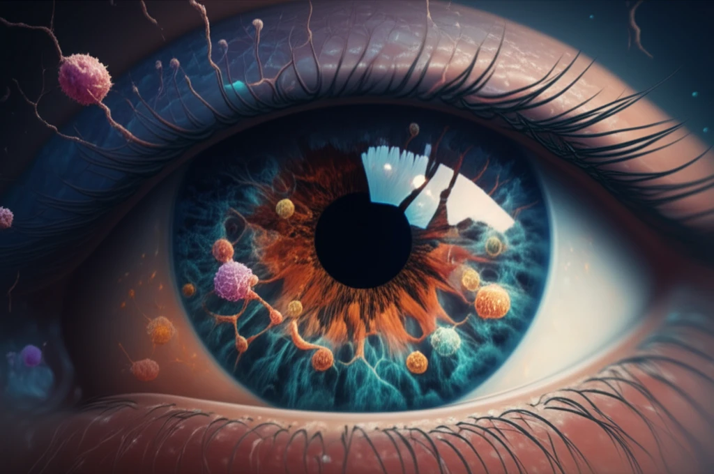
Glaucoma, a leading cause of irreversible blindness, is characterized by the progressive degeneration of retinal ganglion cells (RGCs). While elevated intraocular pressure (IOP) is a known risk factor, many individuals develop glaucoma without high IOP, a condition known as normal-tension glaucoma. This suggests that other factors, including the immune system, may play a significant role in the disease's development.
Emerging research has highlighted the involvement of autoimmunity in glaucoma, with studies identifying elevated levels of autoantibodies against various ocular antigens. Among these antigens, heat shock protein 27 (HSP27) and glial cell line-derived neurotrophic factor (GDNF) have garnered particular attention. However, the precise mechanisms by which these autoantibodies contribute to RGC damage remained unclear, prompting scientists to delve deeper into their effects.
A recent study published in Investigative Ophthalmology & Visual Science explored the impact of GDNF, with or without HSP27, on RGCs and other retinal cells in an autoimmune glaucoma model. This research provides valuable insights into the specific cellular mechanisms involved in glaucoma-related retinal damage, paving the way for the development of targeted therapeutic interventions.
Unraveling the Study: How GDNF and HSP27 Trigger Retinal Damage
Researchers at the University Eye Hospital in Bochum, Germany, conducted a study using a rat model to mimic autoimmune glaucoma. They immunized rats with either GDNF alone or a combination of GDNF and HSP27. The goal was to observe the effects of these immunizations on RGCs and other retinal cells. Over four weeks, the scientists meticulously analyzed the retinas of the immunized animals, employing a range of techniques:
- Immunohistochemistry: Retinal sections were stained with specific markers (Brn-3a, NeuN, GFAP, vimentin, Iba1, ED1, parvalbumin, ChAT, rhodopsin, opsin, PKCa, and recoverin) to identify and quantify different retinal cell types (RGCs, macroglia, microglia, amacrine cells, bipolar cells, and photoreceptors).
- Western Blot Analysis: Protein extracts from the retinas were analyzed to measure the levels of specific proteins associated with RGCs (β-III tubulin), macroglia (GFAP, vimentin), and cholinergic amacrine cells (ChAT).
- Cell Counting and Area Analysis: The number of RGCs, microglia, and amacrine cells was quantified using ImageJ software. The area occupied by GFAP- and vimentin-labeled structures was also measured to assess macroglial activation.
- Intraocular Pressure (IOP) Measurement: IOP was measured using a TonoPen XL to monitor changes in eye pressure.
- Funduscopy: The fundi of the rats were examined to identify any signs of damage or inflammation.
The Implications: New Hope for Glaucoma Therapies
This study illuminates the specific role of GDNF in triggering RGC loss and provides insights into the complex interactions between GDNF and HSP27. The findings suggest that targeting GDNF-mediated autoimmune responses could be a promising therapeutic strategy for glaucoma, especially in cases where IOP is not the primary driver of disease. By understanding the specific antigens that trigger these damaging immune responses, researchers can develop more targeted and effective treatments to protect retinal ganglion cells and prevent vision loss in glaucoma.