Healing stomach lining with glowing threads, symbolizing recovery from gastric perforation.

Gastric Perforation After Endoscopic Ligation: A Rare But Treatable Complication

"Discover how a potentially life-threatening complication of gastric variceal treatment can be managed without surgery, offering hope and insights for patients and practitioners alike."


Variceal bleeding is a serious complication of liver cirrhosis, carrying significant risks despite advancements in treatment. Gastric varices, occurring in a notable percentage of patients with portal hypertension, present unique challenges due to their potential for severe bleeding.

While less frequent than esophageal variceal bleeding, gastric variceal bleeding is often more severe, necessitating substantial blood transfusions and leading to higher rates of rebleeding and mortality. Effective management strategies are crucial to improve patient outcomes.

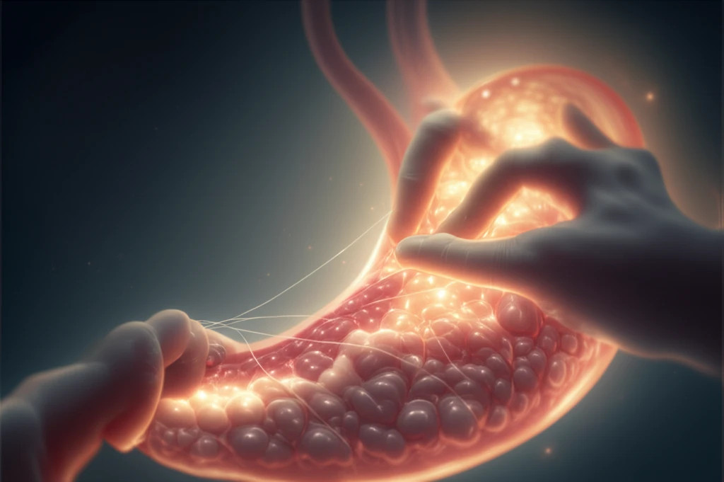
Endoscopic hemostasis, including endoscopic sclerotherapy (ES) and endoscopic variceal ligation (EVL), has become a cornerstone in treating variceal bleeding. EVL, known for its simplicity, is widely used, though complications, especially gastric perforation, are rare. This article delves into a unique case where gastric perforation occurred post-EVL and explores its successful non-surgical management.

Understanding Gastric Perforation After EVL: What Factors Are at Play?

Healing stomach lining with glowing threads, symbolizing recovery from gastric perforation.

In a noteworthy medical case, a 53-year-old male with a history of alcohol use and confirmed liver cirrhosis was admitted to a medical center due to hematemesis. His condition, marked by vital signs indicating distress and physical symptoms such as anemic conjunctiva and abdominal distension, required immediate intervention. Initial laboratory results revealed a hemoglobin level of 7.4 g/dL, a white blood cell count of 11,400/μL, and a platelet count of 67,000/μL, among other findings.

The patient's condition was managed with somatostatin and cefotaxime infusions, complemented by transfusions of packed red blood cells. An urgent esophagogastroduodenoscopy (EGD) was performed, revealing a small non-bleeding esophageal varix and a nodular shaped gastric varix with stigmata at the fundus of the stomach. Subsequently, endoscopic variceal ligation (EVL) was performed to address the bleeding.

After the procedure, the patient developed complications, highlighting critical considerations for patient care and treatment protocols:
  • Two Days Post-EVL: The patient reported abdominal pain, leading to the discovery of a perforation at the post-EVL ulcer base on the gastric fundus via follow-up EGD. Air bubbles were also observed outside the posterior gastric wall of the fundus on an abdominal computed tomography scan.
  • Conservative Treatment: Despite the recommendation for emergency surgery, the patient opted for conservative management, including a nothing-by-mouth order, intensive care unit monitoring, and continuous intravenous antibiotics for ten days.
  • Positive Outcomes: Follow-up EGD after ten days showed healing at the perforation site, allowing for the gradual reintroduction of oral intake. The patient was discharged 25 days later, and a three-month follow-up EGD confirmed complete healing.
This case illuminates the possibility of managing gastric perforation following EVL without surgical intervention. While techniques such as BRTO and TIPS are available, they are often too invasive for general use. Endoscopic hemostasis, including ES and EVL, is typically the recommended initial therapy. The effects of ES and EVL on variceal bleeding have been shown to be similar, with ES sometimes preferred for its lower rebleeding rate.

Key Takeaways: Navigating Gastric Perforation After EVL

While gastric perforation after EVL is rare, this case underscores the importance of vigilance and preparedness. EVL remains a valuable tool for managing variceal bleeding, but it demands careful execution, particularly when addressing fundal varices. Balancing the sucking force and volume during ligation is crucial to avoid unnecessary tissue damage, and a strong consideration should be given to acid-suppressive therapy to prevent iatrogenic ulcers. This case reinforces that conservative management can be a viable option, leading to full recovery and improved patient outcomes.

About this Article -

This article was crafted using a human-AI hybrid and collaborative approach. AI assisted our team with initial drafting, research insights, identifying key questions, and image generation. Our human editors guided topic selection, defined the angle, structured the content, ensured factual accuracy and relevance, refined the tone, and conducted thorough editing to deliver helpful, high-quality information.See our About page for more information.

This article is based on research published under:

DOI-LINK: 10.3346/jkms.2013.28.4.624, Alternate LINK

Title: Spontaneous Healing Of Gastric Perforation After Endoscopic Ligation For Gastric Varices

Subject: General Medicine

Journal: Journal of Korean Medical Science

Publisher: Korean Academy of Medical Sciences

Authors: Jung Ho Kim, Hong Dae Ahn, Kwang An Kwon, Yoon Jae Kim, Jun-Won Chung, Dong Kyun Park, Ju Hyun Kim

Published: 2013-01-01

Everything You Need To Know

1

What are the primary methods for stopping variceal bleeding, and when is each typically used?

The primary methods for stopping variceal bleeding are endoscopic hemostasis techniques, including endoscopic sclerotherapy (ES) and endoscopic variceal ligation (EVL). EVL is often preferred initially due to its simplicity. However, ES might be considered when a lower rebleeding rate is desired, despite having similar effects on initial variceal bleeding control. More invasive procedures like balloon-occluded retrograde transvenous obliteration (BRTO) and transjugular intrahepatic portosystemic shunt (TIPS) are reserved for cases where endoscopic methods are not sufficient due to their invasive nature and potential complications.

2

How does endoscopic variceal ligation (EVL) contribute to the risk of gastric perforation, and what precautions can be taken to minimize this risk?

Endoscopic variceal ligation (EVL) can lead to gastric perforation, though rarely, especially when treating fundal varices. The risk stems from excessive sucking force or volume during ligation, which can damage the tissue. Precautions include carefully balancing the sucking force and volume during the procedure to avoid unnecessary tissue damage. Additionally, administering acid-suppressive therapy can help prevent iatrogenic ulcers at the ligation site, further reducing the risk of perforation.

3

In what situations might conservative management be considered over surgical intervention for gastric perforation after endoscopic variceal ligation (EVL)?

Conservative management, involving measures like a nothing-by-mouth order, intensive care unit monitoring, and continuous intravenous antibiotics, can be considered for gastric perforation following endoscopic variceal ligation (EVL) when the patient's condition is stable and there are no signs of widespread sepsis or peritonitis. This approach allows the perforation site to heal naturally, as demonstrated in the case where the patient showed complete healing after ten days of conservative treatment and was discharged without surgical intervention. However, this decision should be carefully evaluated based on the patient's overall clinical status and response to initial treatment.

4

What are the key differences between endoscopic sclerotherapy (ES) and endoscopic variceal ligation (EVL) in managing variceal bleeding, and how do these differences impact treatment decisions?

Both endoscopic sclerotherapy (ES) and endoscopic variceal ligation (EVL) are effective in managing variceal bleeding. Endoscopic variceal ligation (EVL) is favored for its simplicity and ease of application. Endoscopic sclerotherapy (ES), on the other hand, may be preferred in situations where minimizing the risk of rebleeding is a primary concern, as some studies suggest it has a lower rebleeding rate compared to EVL. The choice between ES and EVL depends on factors such as the patient's specific condition, the location and severity of the varices, and the endoscopist's expertise.

5

Why is gastric variceal bleeding considered more dangerous than esophageal variceal bleeding, and what implications does this have for treatment strategies?

Gastric variceal bleeding is often more severe than esophageal variceal bleeding, necessitating substantial blood transfusions and leading to higher rates of rebleeding and mortality. This increased severity is due to the nature and location of gastric varices, which can be more challenging to access and treat endoscopically. As a result, treatment strategies for gastric variceal bleeding often require a more aggressive and vigilant approach, potentially involving a combination of endoscopic techniques, pharmacological interventions like somatostatin, and close monitoring in an intensive care setting to ensure optimal patient outcomes.

Newsletter Subscribe

Subscribe to get the latest articles and insights directly in your inbox.