Gas Embolism After Lung Biopsy: What You Need to Know
"A rare but serious complication, gas embolism following a lung biopsy requires quick diagnosis and treatment. Here’s how to stay informed and prepared."
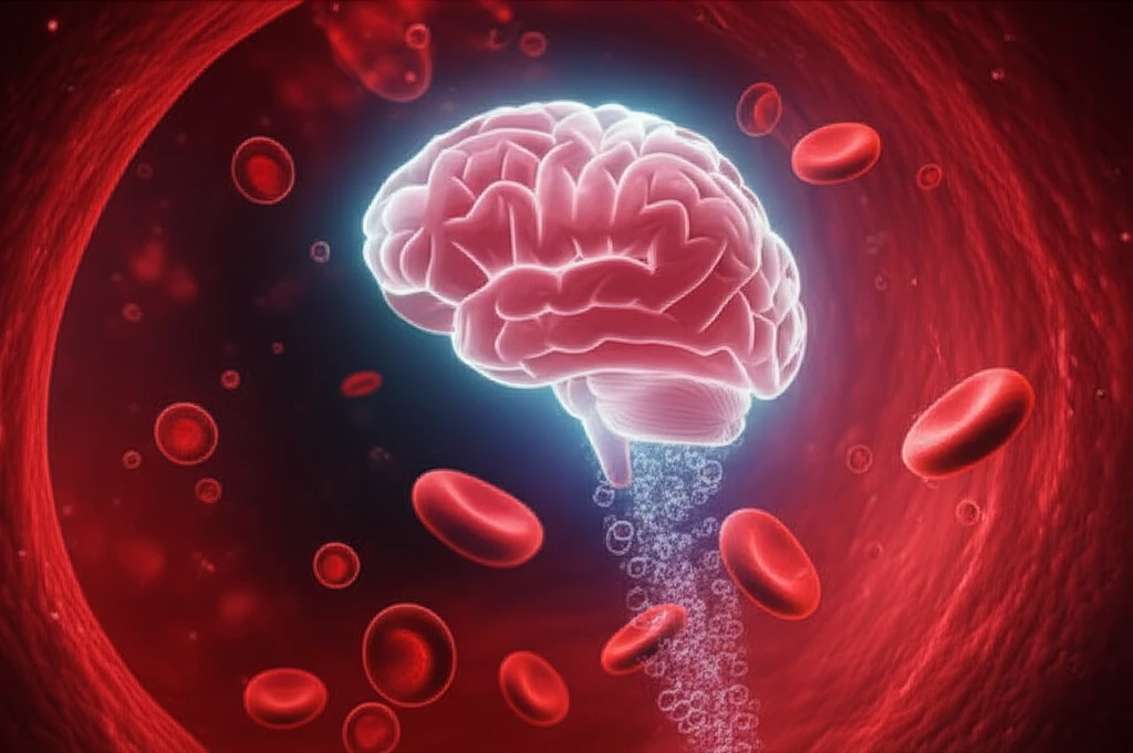
Cerebral ischemia, or stroke, is a rare complication that can occur during surgical or interventional radiology procedures. While uncommon, it's crucial to recognize the potential for such events and understand the various underlying causes.
Gas embolism is one of the possible causes, making swift diagnosis and treatment essential. When air bubbles enter the bloodstream and block blood flow to the brain, the consequences can be severe.
This article explores a case of ischemic stroke following a CT-guided lung biopsy complicated by gas embolism, emphasizing the importance of vigilance and preparedness in medical settings.
Recognizing the Signs: What Does Gas Embolism Look Like?
A 56-year-old male, a smoker with a history of laryngeal cancer treated with radio-chemotherapy, underwent a follow-up chest CT scan. The scan revealed a 17mm pulmonary nodule in the left Fowler segment, prompting a CT-guided lung biopsy.
- Immediate Post-Procedure Symptoms: Sudden loss of consciousness, seizures, and incontinence.
- Neurological Deficits: Weakness on one side of the body, especially affecting the face and arm.
- Imaging Findings: CT scans showing air bubbles in the brain's blood vessels.
Preventing Future Incidents: Steps to Minimize Risk
Gas embolism is a rare but serious complication of transthoracic lung biopsies. Recognizing the signs and acting quickly are essential. Early diagnosis via CT scan and prompt treatment, ideally with hyperbaric oxygen therapy, can significantly improve patient outcomes and reduce the risk of long-term complications.