Gallbladder Surprises: When Medical Scans Go Wrong
"A routine scan revealed a rare case of prolonged contrast material retention, leading to unexpected biliary colic and raising questions about diagnostic accuracy."
Imagine undergoing a routine medical scan only to have it lead to a completely unexpected health issue. This is precisely what happened in a recent case involving a 38-year-old man who initially presented for surgery with a history of postprandial, epigastric pain linked to fatty foods. The twist? His symptoms were eventually traced back to an earlier CT scan where a contrast agent lingered far longer than it should have.
Before his symptoms began, the man had a CT scan of his abdomen and pelvis using oral diatrizoate meglumine and IV iohexol contrast. Initially, the scan pointed toward a small-bowel obstruction without any signs of gallstones. However, as time passed, he developed symptoms consistent with biliary colic—a type of abdominal pain caused by gallstones or a blockage in the bile ducts.
What followed was a diagnostic journey that highlights the complexities of medical imaging and the importance of considering rare complications. This article delves into how retained contrast material mimicked gallstones, leading to diagnostic confusion and ultimately requiring surgical intervention to resolve the man's biliary colic.
The Case of the Lingering Contrast: How a Scan Led to Surgery
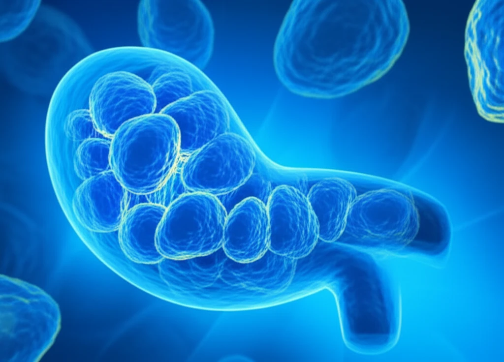
Eighteen months after the initial CT scan, the man underwent another scan using the same contrast material. This time, the images revealed a layering of radio-opaque material within his gallbladder, extending into the cystic duct. To further investigate, a non-contrasted prone CT scan was performed, which showed that the radio-opaque material was dependent, meaning it shifted with gravity. The imaging findings were perplexing, as they resembled an atypical appearance of gallstones.
- Hydropic Fluid: An unusual amount of fluid inside the gallbladder.
- Pigmented Gallstones: Gallstones with a dark color, indicating the presence of bile pigments.
- Clay-like Material: A thick, white substance that resembled clay, which turned out to be the retained contrast material.
Lessons Learned: What This Rare Case Means for You
This unusual case serves as a reminder of the complexities and potential pitfalls of medical imaging. While CT scans and contrast agents are invaluable tools for diagnosis, they are not without risks. The prolonged retention of contrast material, as seen in this case, is a rare but important consideration for clinicians.
For patients, this case underscores the importance of communicating any unusual symptoms or medical history to healthcare providers. If you experience persistent abdominal pain or discomfort after undergoing a CT scan with contrast, it's crucial to inform your doctor, as it could potentially be related to retained contrast material.
Ultimately, this case highlights the need for thorough investigation and consideration of atypical presentations in medical diagnosis. It also reinforces the importance of vigilant post-operative care to ensure complete resolution of symptoms and prevent potential complications.