Heart with flip-flop symbol representing shifting blood flow under stress.

Flip-Flop Heart: When Stress Reveals a Hidden Danger

"Could a reversible perfusion defect in your heart indicate severe coronary artery disease? Learn how a 'flip-flop' effect can be a warning sign."


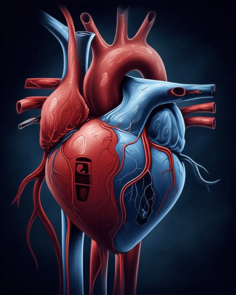
Advancements in gamma cameras and the use of specific tracers have renewed interest in evaluating right ventricular (RV) myocardial perfusion alongside left ventricular assessments. Traditionally, cardiac imaging focuses on the left ventricle, but the right ventricle can offer crucial insights into overall heart health.

A recent case highlights this importance: a patient presented with a reversible perfusion defect in the left anterior descending (LAD) arterial territory. This resulted in a notable visualization of the RV myocardium specifically in post-stress myocardial perfusion imaging (MPI) compared to rest images. This unusual pattern prompted further investigation and revealed a significant underlying issue.

This article will delve into this case, explaining the 'flip-flop' phenomenon observed in myocardial perfusion imaging. We'll explore how this seemingly paradoxical finding can serve as an indirect indicator of severe left ventricular coronary artery disease, offering valuable insights for both patients and healthcare professionals.

The Case: A 'Flip-Flop' Heart

Heart with flip-flop symbol representing shifting blood flow under stress.

A 64-year-old woman, long-time diabetic patient, sought medical help due to worsening chest pain and shortness of breath. Her symptoms had progressed significantly, impacting her daily life. Initial examination revealed abnormalities in heart wall motion, specifically in the region supplied by the left anterior descending artery (LAD).

An electrocardiogram (ECG) showed abnormal ventricular contractions and significant ST-segment depression, indicative of potential heart issues. To further investigate, doctors performed a stress test using adenosine and a special imaging technique called 99mTc-sestamibi gated SPECT/CT MPI.

  • Resting State: The heart appeared to receive adequate blood flow in the LAD territory.
  • Under Stress: A perfusion defect (reduced blood flow) appeared in the LAD territory.
  • RV Visualization: Notably, the right ventricle (RV) was more visible in the post-stress images than in the resting images.
Coronary angiography, a more invasive imaging procedure, confirmed the underlying problem: a complete blockage in the proximal LAD. The heart had developed collateral vessels from the right coronary artery (RCA) to compensate for the blockage, maintaining blood flow at rest. This created a 'flip-flop' effect: blood flow diverted to the RCA under stress, making the RV more prominent in imaging.

What Does This Mean for You?

The case highlights the importance of considering the right ventricle in cardiac imaging. While often overlooked, RV visualization during stress MPI can be a clue to underlying severe left-sided coronary artery disease. The 'flip-flop' phenomenon, where blood flow shifts between the left and right sides of the heart under stress, provides valuable diagnostic information.

This case underscores the heart's remarkable ability to compensate for blockages through collateral vessel formation. However, this compensation isn't always enough, and stress can reveal the underlying limitations. Early detection and intervention, such as bypass grafting, are crucial in managing severe LAD stenosis and preventing further heart damage.

If you experience chest pain or shortness of breath, especially during exertion, it's essential to seek medical attention. Discuss your concerns with your doctor, and ask about appropriate cardiac imaging tests to assess your heart health. Understanding the potential for 'flip-flop' patterns in myocardial perfusion imaging can lead to earlier diagnosis and better outcomes.

About this Article -

This article was crafted using a human-AI hybrid and collaborative approach. AI assisted our team with initial drafting, research insights, identifying key questions, and image generation. Our human editors guided topic selection, defined the angle, structured the content, ensured factual accuracy and relevance, refined the tone, and conducted thorough editing to deliver helpful, high-quality information.See our About page for more information.

This article is based on research published under:

DOI-LINK: 10.1007/s12350-018-01565-z, Alternate LINK

Title: Flip-Flop Right Ventricle Myocardial Perfusion On Stress-Rest 99Mtc-Mibi Myocardial Perfusion Scintigraphy: An Indirect Evidence For Severe Left Ventricular Coronary Arterial Disease?

Subject: Cardiology and Cardiovascular Medicine

Journal: Journal of Nuclear Cardiology

Publisher: Springer Science and Business Media LLC

Authors: Shelvin Kumar Vadi, Saurabh Mehrotra, Ashwani Sood, Madan Parmar, Vaishnavi Dasagrandhi, Komalpreet Kaur, Bhagwant Rai Mittal

Published: 2018-12-13

Everything You Need To Know

1

What is the 'flip-flop' phenomenon in myocardial perfusion imaging, and what does it indicate?

The 'flip-flop' phenomenon in myocardial perfusion imaging refers to a situation where the right ventricle (RV) is more visible in post-stress images compared to resting images. This seemingly paradoxical finding suggests that blood flow is being diverted to the right coronary artery (RCA) under stress, potentially indicating a severe blockage in the left anterior descending artery (LAD). It is an indirect indicator of severe left ventricular coronary artery disease.

2

What symptoms and test results led to the discovery of the 'flip-flop' heart condition in the described patient?

In the case described, the patient experienced worsening chest pain and shortness of breath. An electrocardiogram (ECG) showed abnormal ventricular contractions and significant ST-segment depression. A stress test using adenosine and 99mTc-sestamibi gated SPECT/CT MPI revealed a perfusion defect in the left anterior descending artery (LAD) territory under stress and notable visualization of the right ventricle (RV). Coronary angiography confirmed a complete blockage in the proximal LAD, with collateral vessels from the right coronary artery (RCA) compensating for blood flow at rest.

3

How have advancements in cardiac imaging technology improved the detection and understanding of conditions like the 'flip-flop' phenomenon?

Advancements in gamma cameras and the use of specific tracers have enabled better evaluation of the right ventricular (RV) myocardial perfusion alongside left ventricular assessments. This is crucial because the right ventricle can offer important insights into overall heart health, including indications of severe coronary artery disease on the left side, as indicated by the 'flip-flop' phenomenon.

4

What does it mean when a reversible perfusion defect is found in the left anterior descending (LAD) artery, and how does it relate to the 'flip-flop' phenomenon?

When a reversible perfusion defect is observed in the left anterior descending (LAD) artery territory, it means that while there is adequate blood flow at rest, blood flow becomes restricted or reduced during stress. This can cause the 'flip-flop' phenomenon, where the right ventricle (RV) becomes more visible in post-stress myocardial perfusion imaging (MPI). This phenomenon suggests that there may be severe underlying coronary artery disease and can indicate a blockage in the LAD, which could be managed via intervention or medication.

5

Why is it important to assess the right ventricle (RV) in cardiac imaging, and how does it connect to conditions affecting the left ventricle?

While cardiac imaging traditionally focuses on the left ventricle, evaluating the right ventricle (RV) provides crucial insights. The 'flip-flop' phenomenon, observed through myocardial perfusion imaging (MPI), shows that increased RV visualization under stress can be a sign of severe left-sided coronary artery disease, specifically in the left anterior descending artery (LAD). This highlights the interconnectedness of the heart's chambers and the importance of considering both ventricles in diagnosis to fully assess and address cardiac health.

Newsletter Subscribe

Subscribe to get the latest articles and insights directly in your inbox.