Advanced skin imaging technology revealing healthy skin cells and vessels.

Flawless Photos: How Motion Correction Revolutionizes Skin Imaging

"Discover how a groundbreaking algorithm is eliminating blur and distortion in optoacoustic mesoscopy, paving the way for clearer, more accurate skin health assessments."


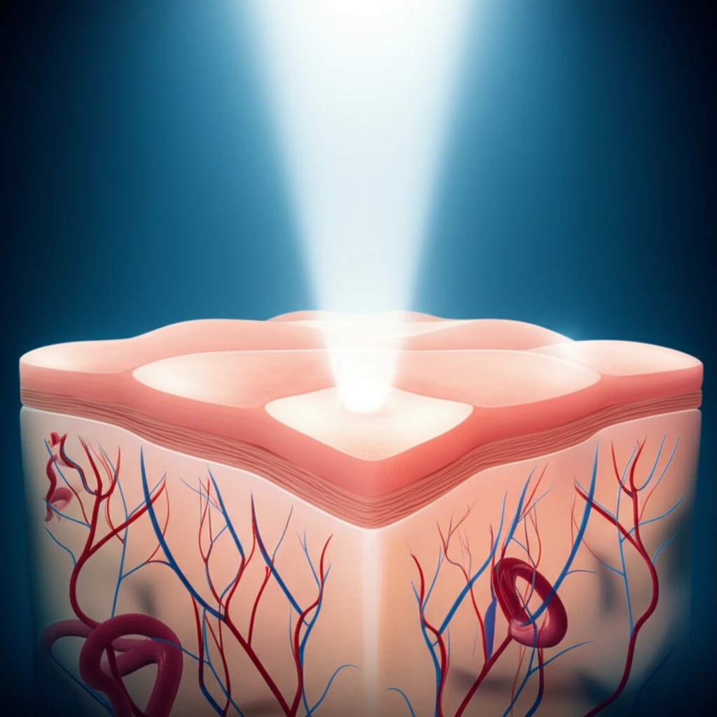
Imagine trying to take a photo while your subject is constantly moving. The result? A blurry, distorted mess. This is a common challenge in medical imaging, particularly when examining delicate tissues like skin. Traditional methods often struggle to produce clear, accurate images due to the patient's subtle movements – breathing, twitching, or even slight shifts in position.

But what if there was a way to freeze that motion, digitally? That's precisely what a team of researchers has achieved with a revolutionary motion correction algorithm designed for optoacoustic mesoscopy (RSOM). This innovative technique promises to eliminate blur and distortion, providing clinicians with unprecedented clarity when assessing skin health.

Optoacoustic mesoscopy, also known as photoacoustic mesoscopy, is a powerful imaging technique that provides novel insights into vascular morphology and pathophysiological biomarkers of skin inflammation. RSOM uses ultra-wideband detection and focused ultrasound transducers, achieving high resolution at depths that other optical imaging methods cannot reach. Until now, motion artifacts have limited its effectiveness, but this new algorithm changes everything.

The Science Behind the Stillness: How the Motion Correction Algorithm Works

Advanced skin imaging technology revealing healthy skin cells and vessels.

The heart of this innovation lies in how the algorithm analyzes disruptions in the ultrasound wave front. Think of it like ripples in a pond – any disturbance changes the pattern. In this case, the algorithm observes how the vertical movement of melanin (the pigment in skin) disrupts these waves during the RSOM scan.

Here’s a breakdown of the process:

  • Mapping the Disruption: The algorithm first identifies distortions in the ultrasound wave front caused by skin movement.
  • Creating a Synthetic Surface: From this disrupted surface, the algorithm generates a smooth, synthetic surface representing the ideal, motionless skin.
  • Calculating the Offset: The difference between the real, disrupted surface and the synthetic surface provides a precise measurement of how much the skin moved during the scan.
  • Correcting the Image: This offset is then used to correct the relative position of the ultrasound detector, effectively 'undoing' the motion and producing a clear image.
The result is a significantly sharper, more accurate image, allowing clinicians to see details they might have missed before. The technology has the potential to enhance diagnoses and treatment monitoring of skin conditions.

The Future of Skin Diagnostics: RSOM and Beyond

This motion correction algorithm is not just a technical achievement; it's a significant step forward for clinical dermatology. By eliminating motion artifacts, the algorithm unlocks the full potential of RSOM, enabling more accurate diagnoses, better treatment monitoring, and a deeper understanding of skin health. This breakthrough paves the way for a new era of non-invasive skin diagnostics, potentially transforming how we detect and manage a wide range of dermatological conditions.

About this Article -

This article was crafted using a human-AI hybrid and collaborative approach. AI assisted our team with initial drafting, research insights, identifying key questions, and image generation. Our human editors guided topic selection, defined the angle, structured the content, ensured factual accuracy and relevance, refined the tone, and conducted thorough editing to deliver helpful, high-quality information.See our About page for more information.

Everything You Need To Know

1

What is optoacoustic mesoscopy and how does it relate to skin imaging?

Optoacoustic mesoscopy, also known as photoacoustic mesoscopy, is a powerful imaging technique used to visualize skin in detail. It provides insights into the vascular morphology and pathophysiological biomarkers of skin inflammation. RSOM utilizes ultra-wideband detection and focused ultrasound transducers. This method is crucial for assessing skin health and diagnosing various dermatological conditions.

2

How does the motion correction algorithm work in optoacoustic mesoscopy?

The motion correction algorithm analyzes disruptions in the ultrasound wave front during RSOM scans. First, it maps distortions caused by skin movement. Then, it creates a synthetic surface representing motionless skin. By calculating the offset between the real, disrupted surface and the synthetic one, the algorithm measures skin movement. Finally, it corrects the image by adjusting the relative position of the ultrasound detector to eliminate motion artifacts and produce a clear image.

3

What are the limitations of traditional skin imaging methods, and how does RSOM overcome them?

Traditional methods often struggle to produce clear skin images due to patient movement, which causes blur and distortion. RSOM overcomes these limitations by employing a motion correction algorithm. This algorithm digitally 'freezes' the motion, allowing clinicians to obtain sharp and accurate images, thus improving the assessment of skin health and the diagnosis of skin conditions.

4

What are the implications of using RSOM with the new motion correction algorithm for clinical dermatology?

The integration of RSOM with the motion correction algorithm represents a significant advancement for clinical dermatology. It enables more accurate diagnoses, better treatment monitoring, and a deeper understanding of skin health. The technology has the potential to transform how dermatological conditions are detected and managed, paving the way for non-invasive skin diagnostics.

5

Can you explain the specific steps the algorithm takes to correct motion artifacts in RSOM?

The motion correction algorithm follows these steps: First, it maps distortions in the ultrasound wave front caused by skin movement. Second, it generates a smooth, synthetic surface representing motionless skin. Third, it calculates the offset by determining the difference between the real, disrupted surface and the synthetic surface. Lastly, it uses this offset to correct the image by adjusting the relative position of the ultrasound detector, effectively undoing the motion and producing a clear image.

Newsletter Subscribe

Subscribe to get the latest articles and insights directly in your inbox.