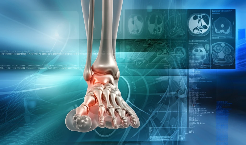
Flat Feet, Big Problems? Unlocking the Secrets to Hallux Valgus and First Ray Instability
"New insights into adult-acquired flatfoot deformity (AAFD) and its link to hallux valgus, offering a clearer understanding for patients and professionals alike."
Do you ever feel like your feet are the foundation of, well, everything? Turns out, that feeling isn't too far off. Our feet play a crucial role in our overall alignment and movement. When things go awry down there, it can lead to a cascade of issues, particularly for those with adult-acquired flatfoot deformity (AAFD). AAFD, often characterized by the collapse of the arch, doesn't just affect the foot; it's intricately linked to the development of hallux valgus, more commonly known as bunions.
Hallux valgus, that prominent bump at the base of the big toe, isn't merely a cosmetic concern. It signifies an underlying instability in the foot's structure. Traditionally, diagnosing these instabilities has relied on standard X-rays. However, a groundbreaking approach using weightbearing CT imaging (WBCT) is changing the game, offering a three-dimensional look at how the foot functions under the pressure of our body weight. This advanced imaging technique provides a more accurate and comprehensive evaluation of dynamic deformities like AAFD and hallux valgus.
A recent study published in Foot & Ankle Orthopaedics sheds light on the correlation between hallux valgus severity and various foot collapse indicators, all thanks to WBCT measurements. The researchers hypothesized that the flattening of the longitudinal arch, increased hindfoot valgus (a tilting of the heel), and forefoot abduction (where the front of the foot turns outward) would correlate with greater intermetatarsal and hallux valgus angles. Let’s dive into what they discovered and what it means for you.
The Flatfoot-Hallux Valgus Connection: What the WBCT Scans Reveal

In this retrospective comparative study, researchers analyzed WBCT scans from 108 patients diagnosed with stage II AAFD. The group consisted of 36 men and 72 women, with an average age of 54.4 years. Two independent, board-certified orthopedic surgeons, blinded to patient data, meticulously evaluated these scans, focusing on key variables that indicate the severity of hallux valgus and flatfoot deformities. These variables included the 1-2 intermetatarsal angle (the angle between the first and second metatarsals), the hallux valgus angle (measuring the bunion's severity), and other crucial alignment measurements.
- Both the intermetatarsal (IM) and hallux valgus (HV) angles showed a strong correlation with each other (p<0.0001).
- Increased IM angle correlated with increased talocalcaneal angle (26.0°±10.3°, p<0.0001), talus-first metatarsal angle (19.0°±13.6°, p=0.0001), and hindfoot alignment angles (22.3°±12.9°, p= 0.0049). This essentially means that as the flatfoot deformity worsens, the angle between the metatarsals increases, exacerbating the bunion.
- The HV angle correlated with medial cuneiform-floor distance (15.1mm±5.5mm, p=0.0183), talus-first metatarsal angle in both the axial plane (p=0.0004) and sagittal plane (15.7°±8.8°, p=0.0351), and talonavicular uncoverage angle (17.8°±13.9°, p=0.0035). In other words, a more severe bunion is associated with specific changes in the alignment and positioning of bones in the foot.
- Interestingly, hindfoot moment arm and navicular-floor distance were the only AAFD measurements that did not correlate with IM or HV angles. This suggests that these particular measurements might be influenced by other factors or represent different aspects of the deformity.
What Does This Mean for Your Feet?
This study reinforces the idea that flat feet and bunions are often interconnected. By using weightbearing CT imaging, doctors can now gain a more detailed understanding of the complexities of these conditions. If you're experiencing foot pain, have flat feet, or notice a bunion developing, it’s essential to seek a comprehensive evaluation from a foot and ankle specialist. Early diagnosis and appropriate management can help prevent the progression of these deformities and improve your overall quality of life. While this study highlights correlations, it’s crucial to remember that cause and effect can't be definitively determined. However, the findings provide valuable insights that foot and ankle surgeons should consider when evaluating and planning surgical interventions for patients with AAFD.
