CT scan image showing a fish bone perforating the small intestine, leading to peritonitis.

Fish Bone Perforation: A Rare Cause of Peritonitis

"When dinner fights back: A case study exploring how a seemingly harmless fish bone led to a serious abdominal infection and the surgical steps taken to resolve it."


Diagnosing a small bowel perforation caused by a foreign object before surgery is incredibly difficult. These perforations often occur near the ileocecal region, presenting symptoms similar to appendicitis or even generalized peritonitis. In rare instances, these perforations can heal on their own.

This case report details a 70-year-old patient with no significant medical history who was admitted to the emergency department with diffuse abdominal pain that had been worsening for two days. She also experienced some vomiting, but no other accompanying symptoms.

Upon examination, the patient had a fever of 38.6°C but was hemodynamically stable. Her abdomen showed generalized guarding, indicating peritonitis. The rest of her physical examination was unremarkable. Lab results revealed a high white blood cell count of 12,300/mm3 and a C-reactive protein (CRP) level of 94. An initial abdominal X-ray showed no abnormalities.

Unraveling the Diagnosis: How Imaging Revealed the Culprit

CT scan image showing a fish bone perforating the small intestine, leading to peritonitis.

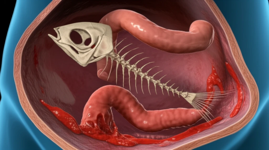
Due to the inconclusive X-ray results, an abdominal CT scan was performed. The scan revealed localized thickening of the small bowel, with regular and symmetrical circumferential involvement. There was also enhancement of the mucosa and edema in the submucosa. Within this area, a foreign body, suspected to be a fish bone, appeared to be piercing the intestinal wall, accompanied by a moderate amount of fluid in the peritoneal cavity.

Based on these findings, a diagnosis of peritonitis due to small bowel perforation by a foreign body was made. The decision was made to proceed with surgical intervention.

  • Preoperative Diagnosis Challenges: Highlighting the difficulties in diagnosing small bowel perforations caused by foreign objects, emphasizing the similarity to other abdominal conditions.
  • Importance of Advanced Imaging: Illustrating how a CT scan was crucial in identifying the fish bone and the resulting perforation, leading to a definitive diagnosis.
  • Surgical Intervention: Describing the surgical procedure, including the extraction of the fish bone and the subsequent repair of the perforation.
During surgery, turbid peritoneal fluid was observed, along with a pinpoint perforation in the small bowel. The fish bone was carefully extracted through a small enterotomy, and the perforation was sutured closed. The abdominal cavity was then thoroughly washed and drained. The patient's postoperative recovery was uneventful. She was seen for follow-up appointments three and six months later, and her clinical examination remained normal.

Key Takeaways: Learning from a Rare Case

This case underscores the importance of considering foreign body ingestion in patients presenting with unexplained abdominal pain and peritonitis, even in the absence of a clear history. Advanced imaging techniques like CT scans are invaluable in diagnosing such cases, allowing for prompt surgical intervention and favorable outcomes.

About this Article -

This article was crafted using a human-AI hybrid and collaborative approach. AI assisted our team with initial drafting, research insights, identifying key questions, and image generation. Our human editors guided topic selection, defined the angle, structured the content, ensured factual accuracy and relevance, refined the tone, and conducted thorough editing to deliver helpful, high-quality information.See our About page for more information.

This article is based on research published under:

DOI-LINK: 10.11604/pamj.2013.15.107.3025, Alternate LINK

Title: Péritonite Par Perforation Grêlique Secondaire À Une Arête De Poisson

Subject: General Medicine

Journal: Pan African Medical Journal

Publisher: Pan African Medical Journal

Authors: Karim Ibn Majdoub, Imane Toughrai

Published: 2013-01-01

Everything You Need To Know

1

What is peritonitis and how was it related to this case?

Peritonitis is a serious medical condition characterized by inflammation of the peritoneum, the lining of the abdominal cavity. This patient developed peritonitis due to a small bowel perforation caused by a fish bone. The fish bone pierced the small intestine, allowing bacteria to leak into the abdominal cavity, leading to the inflammation and infection characteristic of peritonitis. Early diagnosis and surgical intervention are crucial to prevent life-threatening complications.

2

Why is it difficult to diagnose a small bowel perforation?

Diagnosing a small bowel perforation caused by a foreign object can be challenging due to the vague and non-specific nature of the symptoms. The symptoms can mimic other conditions like appendicitis. In this case, initial X-ray results were inconclusive. However, the CT scan proved crucial in identifying the fish bone and the resulting perforation, enabling the medical team to make a definitive diagnosis. Advanced imaging, like the CT scan, plays a vital role in such cases, providing detailed images that can reveal the underlying cause of the patient's symptoms.

3

What were the initial signs and symptoms the patient exhibited?

A 70-year-old patient presented with diffuse abdominal pain and other symptoms. Upon examination, the patient exhibited signs of peritonitis, including generalized guarding and fever. The patient's lab results indicated a high white blood cell count and an elevated C-reactive protein (CRP) level, both of which are indicative of an inflammatory response. These findings, along with the patient's symptoms, led to further investigations, including an abdominal CT scan.

4

What surgical steps were taken to address the problem?

Surgical intervention was necessary to address the small bowel perforation caused by the fish bone. The surgical procedure involved extracting the fish bone through a small enterotomy. The perforation in the small bowel was then carefully sutured closed. The abdominal cavity was thoroughly washed and drained to remove any remaining contaminants and minimize the risk of infection. This surgical approach successfully resolved the perforation and allowed the patient to recover.

5

What key lessons can we learn from this case?

The case underscores the importance of considering foreign body ingestion in patients presenting with unexplained abdominal pain and peritonitis, even without a clear history of ingestion. Advanced imaging techniques, like CT scans, are invaluable in diagnosing such cases. The successful outcome highlights the importance of early diagnosis through imaging and prompt surgical intervention in cases of small bowel perforation due to foreign bodies. This prompt action is key to minimizing complications and ensuring a favorable patient outcome.

Newsletter Subscribe

Subscribe to get the latest articles and insights directly in your inbox.