Surreal illustration of facial nerve protection during ear surgery

Facial Canal Dehiscence: Are You at Risk? What You Need to Know

"Understanding the causes, risks, and prevention of facial nerve damage during chronic otitis media surgery."


Imagine undergoing ear surgery only to face an unexpected complication: facial paralysis. This is a real concern for otolaryngologists (ENT surgeons) and patients dealing with chronic otitis media (COM), an ongoing ear infection. Facial nerve paralysis doesn't just affect physical appearance; it can lead to social isolation and a hit to one's self-esteem.

The culprit behind this complication can be a condition called Facial Canal Dehiscence (FCD). FCD refers to an absence of bone covering the facial nerve canal, leaving the nerve vulnerable during surgery. While a solid understanding of anatomy is crucial, surgeons also need to be aware of the possibility of FCD during ear operations to prevent nerve damage.

A recent study investigated the rate and location of FCD in patients undergoing surgery for chronic otitis media, both with and without cholesteatoma (a skin cyst in the middle ear). By understanding the prevalence and common locations of FCD, surgeons can take extra precautions to protect the facial nerve.

What is Facial Canal Dehiscence (FCD) and Why Does It Matter?

Surreal illustration of facial nerve protection during ear surgery

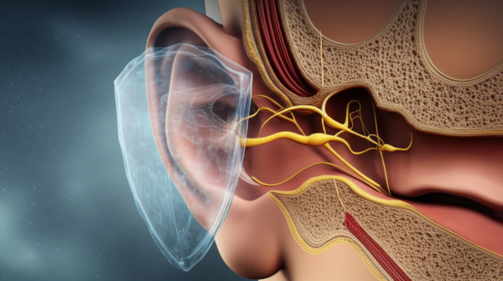
Facial Canal Dehiscence (FCD) is a condition where the bony covering of the facial nerve canal is missing. This leaves the facial nerve exposed and vulnerable to injury during ear surgeries. FCD can be present from birth or develop over time due to bone erosion caused by chronic ear infections, particularly those involving cholesteatoma.

The study analyzed data from 372 patients who underwent mastoidectomy (a procedure to remove infected air cells in the mastoid bone) for chronic otitis media. The research focused on identifying instances of FCD observed during the operations. Key findings include:

  • Prevalence: FCD was found in 11.29% of the patients.
  • Association with Cholesteatoma: FCD was more common in patients with cholesteatoma (88.1%) compared to those without (11.9%).
  • Location: The most frequent location of FCD was in the tympanic segment (76.19%) of the facial canal, which is near the oval window.
  • Ossicular Erosion: In cases where FCD was present, erosion of all three ossicles (small bones in the middle ear) was statistically more frequent.
These findings highlight the importance of being aware of FCD, especially when dealing with chronic otitis media and cholesteatoma. The location of the dehiscence and the degree of ossicular erosion can help surgeons anticipate the risk and take necessary precautions.

Protecting the Facial Nerve: What Surgeons Can Do

The study emphasizes the need for surgeons to be extra vigilant when operating on patients with chronic otitis media, especially those with cholesteatoma. Given that FCD is most commonly found near the oval window, careful examination of this area is crucial.

The study suggests that if the stapes (one of the ossicles) or all three ossicles are eroded, the risk of FCD is higher. Surgeons should exercise extreme caution in these situations to minimize the risk of facial nerve injury.

While preoperative imaging, such as micro-CT scans, can help identify FCD, the ultimate determination often relies on careful intraoperative examination. By staying informed about the prevalence, location, and associated factors of FCD, surgeons can enhance their ability to protect the facial nerve and improve patient outcomes.

About this Article -

This article was crafted using a human-AI hybrid and collaborative approach. AI assisted our team with initial drafting, research insights, identifying key questions, and image generation. Our human editors guided topic selection, defined the angle, structured the content, ensured factual accuracy and relevance, refined the tone, and conducted thorough editing to deliver helpful, high-quality information.See our About page for more information.

This article is based on research published under:

DOI-LINK: 10.1007/s00405-018-5198-7, Alternate LINK

Title: Facial Canal Dehiscence Rate: A Retrospective Analysis Of 372 Chronic Otitis Media Cases

Subject: General Medicine

Journal: European Archives of Oto-Rhino-Laryngology

Publisher: Springer Science and Business Media LLC

Authors: M. Tayyar Kalcioglu, Osman Kilic, Ozan Tuysuz, Serkan Serifler, Muhammet Tekin

Published: 2018-11-12

Everything You Need To Know

1

What is Facial Canal Dehiscence (FCD), and why is it a concern for those undergoing ear surgery?

Facial Canal Dehiscence (FCD) is a condition where the bone protecting the facial nerve canal is missing, leaving the Facial nerve vulnerable. In the context of ear surgery, particularly for chronic otitis media (COM), the presence of FCD significantly increases the risk of damaging the Facial nerve during the procedure. This can lead to Facial nerve paralysis, which has profound implications for a patient's physical appearance, social interactions, and self-esteem.

2

What is the Facial nerve, and why is it so important?

The Facial nerve is a cranial nerve that controls facial expressions. The Facial nerve travels through the Facial nerve canal within the temporal bone of the skull. If the canal is open, the Facial nerve is exposed and at a higher risk of damage. The significance here is that damage to the Facial nerve during ear surgery, often performed for chronic otitis media (COM), can result in facial paralysis. The implications of this can range from difficulties in everyday functions, such as eating and closing the eye, to psychological distress.

3

How does chronic otitis media (COM) relate to Facial Canal Dehiscence (FCD) and facial nerve damage?

Chronic otitis media (COM) is a persistent or recurring infection of the middle ear. In the context provided, COM is a significant factor because it can lead to the development of Facial Canal Dehiscence (FCD). The infection can erode the bone that protects the Facial nerve canal. Furthermore, the study indicates that FCD is more common in patients with COM, especially when complicated by cholesteatoma, increasing the risk of Facial nerve damage during surgical intervention. The implications are higher surgical risk and the potential for serious complications, like facial paralysis.

4

Why is the location of Facial Canal Dehiscence (FCD) near the oval window significant?

The oval window is an opening in the bony wall of the middle ear, which connects to the inner ear. The study shows that the most frequent location of Facial Canal Dehiscence (FCD) is near the oval window. Therefore, surgeons need to be particularly careful when operating in this area, as the exposed Facial nerve is at high risk of injury. The implications of this finding are that surgeons must pay special attention to the tympanic segment, which is near the oval window, during surgical procedures to prevent inadvertent damage to the Facial nerve.

5

How does cholesteatoma affect the risk of Facial nerve damage during surgery?

Cholesteatoma is an abnormal skin cyst that develops in the middle ear. The study highlights that Facial Canal Dehiscence (FCD) is more common in patients with cholesteatoma than those without. This association underscores the need for surgeons to be even more cautious when operating on patients who have cholesteatoma. This is because the presence of cholesteatoma indicates an increased risk of Facial nerve damage due to the higher likelihood of FCD. The implications are that these patients may require more complex surgical approaches and extra care during surgery to mitigate the risk of Facial nerve injury.

Newsletter Subscribe

Subscribe to get the latest articles and insights directly in your inbox.