Illustration of liver displacement due to diaphragmatic eventration.

Everted Liver: A Rare Twist in Diaphragmatic Eventration

"Unveiling a unique case of liver displacement secondary to a diaphragmatic anomaly."


Diaphragmatic eventration, a condition characterized by abnormal elevation of the diaphragm, typically presents with respiratory or gastrointestinal symptoms. However, a recent case highlights an unusual consequence of this condition: an everted liver. This article delves into the details of this rare presentation, drawing from a published case report to shed light on its diagnosis and management.

The original research, documented in 'Clinical Gastroenterology and Hepatology,' presents the case of a 67-year-old male with a history of chronic obstructive bronchopneumopathy who presented with thoracic pain and cough. Investigations revealed an unexpected finding: the liver had become displaced, a condition termed 'everted liver,' due to diaphragmatic eventration.

This article aims to break down the complexities of this case, explaining the underlying mechanisms of diaphragmatic eventration and its potential impact on abdominal organs. We'll explore the diagnostic process, the specific findings in this patient, and the rationale behind the conservative management approach.

What is Diaphragmatic Eventration?

Illustration of liver displacement due to diaphragmatic eventration.

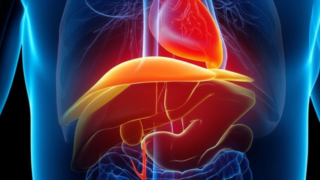
Diaphragmatic eventration refers to an abnormal elevation of part or all of the diaphragm. Unlike diaphragmatic hernias, where there is a tear or opening in the diaphragm allowing abdominal organs to protrude into the chest cavity, eventration involves a weakening or thinning of the diaphragmatic muscle itself. This weakened area bulges upward, displacing the structures above it.

The causes of diaphragmatic eventration can vary. Some cases are congenital, meaning they are present at birth, often due to incomplete muscular development of the diaphragm. Acquired eventration can result from phrenic nerve paralysis (which controls diaphragm movement), trauma, or surgery. In the case presented, the eventration was attributed to atrophy (wasting) of the diaphragmatic muscle fibers, potentially leading to phrenic nerve paralysis.

  • Chest X-ray: Visualizes the elevated diaphragm.
  • CT Scan: Provides detailed images of the diaphragm and surrounding structures, helping to confirm the diagnosis and identify any associated abnormalities.
In this specific case, a 67-year-old man presented with thoracic pain and cough. A chest radiograph revealed an elevated right hemidiaphragm. Subsequent CT scanning confirmed right diaphragmatic eventration. Notably, the liver and gallbladder were displaced downwards, positioned just beneath the right hemidiaphragm, a finding consistent with liver eversion.

Living with Diaphragmatic Eventration

The management of diaphragmatic eventration depends largely on the severity of symptoms. Many individuals with eventration experience no symptoms and require no specific treatment. In cases where symptoms such as shortness of breath or gastrointestinal issues are present, various interventions may be considered.

In the reported case, the patient was asymptomatic despite the unusual liver displacement. As a result, the medical team opted for conservative management, involving observation and supportive care. This approach highlights that not all cases of diaphragmatic eventration require invasive intervention.

This case serves as a reminder of the diverse ways in which diaphragmatic eventration can manifest. While liver eversion is a rare occurrence, it underscores the importance of considering anatomical variations when evaluating patients with chest or abdominal complaints. Further research and case studies will help to better understand the long-term implications of this unusual condition and refine management strategies.

About this Article -

This article was crafted using a human-AI hybrid and collaborative approach. AI assisted our team with initial drafting, research insights, identifying key questions, and image generation. Our human editors guided topic selection, defined the angle, structured the content, ensured factual accuracy and relevance, refined the tone, and conducted thorough editing to deliver helpful, high-quality information.See our About page for more information.

Everything You Need To Know

1

What exactly is diaphragmatic eventration, and how does it differ from a diaphragmatic hernia?

Diaphragmatic eventration describes an abnormal elevation of the diaphragm, where part or all of the muscle is weakened, causing it to bulge upwards. Unlike a diaphragmatic hernia, where there's a tear allowing organs to protrude, eventration involves the thinning, not a tear, of the diaphragm muscle. The causes can be congenital, due to incomplete muscle development, or acquired, resulting from phrenic nerve paralysis, trauma, or surgery. The diaphragm's inability to contract and descend properly leads to reduced space in the chest cavity and potential displacement of abdominal organs.

2

What does "everted liver" mean in the context of diaphragmatic eventration?

Everted liver, in the context of diaphragmatic eventration, refers to the displacement of the liver from its normal anatomical position due to the abnormal elevation of the diaphragm. The weakened diaphragm bulges upwards, pushing the liver downwards and causing it to become 'everted'. This condition is often discovered incidentally during imaging for other symptoms, such as thoracic pain or cough, as was the case with the 67-year-old patient in the 'Clinical Gastroenterology and Hepatology' case report.

3

How was diaphragmatic eventration with everted liver diagnosed in the 67-year-old patient?

In this specific case, the patient, a 67-year-old male, presented with thoracic pain and cough. Diagnostic imaging, including a chest X-ray and CT scan, revealed an elevated right hemidiaphragm indicative of diaphragmatic eventration. What was particularly notable was the downward displacement of the liver and gallbladder, positioned just beneath the right hemidiaphragm, confirming the everted liver. The diaphragmatic eventration was attributed to atrophy of the diaphragmatic muscle fibers, potentially leading to phrenic nerve paralysis.

4

What are the treatment options for diaphragmatic eventration, and why was a conservative approach chosen for the patient with everted liver?

The management approach for diaphragmatic eventration varies depending on the severity of symptoms. Many individuals with eventration are asymptomatic and require no specific treatment. However, when symptoms such as shortness of breath or gastrointestinal issues arise, interventions may be considered. In the specific case of the 67-year-old patient with everted liver, a conservative management approach was chosen. The reasoning behind conservative management often involves monitoring the patient's symptoms and providing supportive care, rather than immediate surgical intervention, especially if the patient's symptoms are mild or stable. Surgical plication can be considered for those with severe symptoms.

5

Besides X-rays and CT scans, what other diagnostic tests can help evaluate diaphragmatic eventration and its impact on respiratory function?

While imaging techniques like chest X-rays and CT scans are valuable for diagnosing diaphragmatic eventration and related conditions such as everted liver, other diagnostic modalities exist. Fluoroscopy, for instance, can assess diaphragm movement, while ultrasound may provide additional visualization. Furthermore, pulmonary function tests can evaluate the impact of diaphragmatic eventration on respiratory function, which wasn't explicitly mentioned in the case but is a relevant consideration. Understanding the degree of respiratory compromise can help guide management decisions and determine the need for interventions beyond conservative measures.

Newsletter Subscribe

Subscribe to get the latest articles and insights directly in your inbox.