Microscopic exosomes illuminated by Raman Spectroscopy.

Early Detection of Pancreatic Cancer: Can Exosomes and Raman Spectroscopy Change the Game?

"A new study explores how analyzing exosomes with surface-enhanced Raman spectroscopy could revolutionize early pancreatic cancer detection, offering hope for improved outcomes."


Pancreatic cancer remains one of the deadliest forms of cancer, primarily because it's often diagnosed at advanced stages. The lack of early, reliable detection methods means that treatments are frequently less effective, and survival rates are tragically low. Finding ways to diagnose pancreatic cancer sooner is a critical challenge for medical researchers.

Exosomes, tiny vesicles released by cells, have emerged as potential biomarkers for various diseases, including cancer. These exosomes contain a wealth of information about the cells they originate from, offering a non-invasive way to peek into the body's processes. However, unlocking the secrets held within these exosomes has been a complex task.

Now, a groundbreaking study has explored the use of surface-enhanced Raman spectroscopy (SERS) to analyze exosomes and detect pancreatic cancer at an early stage. This innovative approach combines the power of nanotechnology with advanced analytical techniques, offering a promising new avenue for early detection and improved patient outcomes.

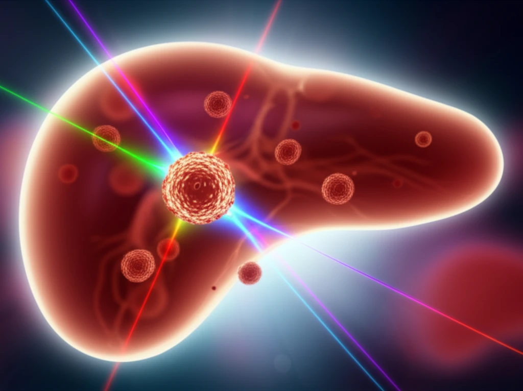
What is Surface-Enhanced Raman Spectroscopy (SERS) and How Does It Work?

Microscopic exosomes illuminated by Raman Spectroscopy.

Raman spectroscopy is a technique that analyzes how light interacts with molecules. When light hits a molecule, it scatters in different ways, creating a unique spectral fingerprint. Surface-enhanced Raman spectroscopy (SERS) amplifies this signal by using metallic nanoparticles, making it possible to detect even trace amounts of specific molecules. This amplification is crucial for analyzing the small amounts of material found in exosomes.

In the context of cancer detection, SERS can identify unique molecular signatures associated with cancer cells. By analyzing the Raman spectra of exosomes, researchers can potentially distinguish between healthy cells and cancerous cells, providing a basis for early diagnosis. The beauty of SERS lies in its label-free nature; it doesn't require specific markers or probes, offering a broad and unbiased view of the exosomal content.

  • Sample Preparation: Exosomes are isolated from blood or cell culture samples.
  • Nanoparticle Enhancement: Exosomes are mixed with metallic nanoparticles (usually gold or silver).
  • Raman Analysis: The sample is illuminated with a laser, and the scattered light is analyzed.
  • Spectral Analysis: The resulting Raman spectrum is analyzed to identify molecular signatures.
The study detailed in the research paper used gold nanoparticles to enhance the Raman signal from exosomes. Researchers coated microscope slides with gold and then deposited gold nanoparticles onto the slides. This process amplified the Raman signals, making the detection of subtle molecular differences easier and more reliable. This enhancement is critical due to the inherently weak Raman scattering efficiency.

The Future of Pancreatic Cancer Detection

This study offers a promising glimpse into the future of pancreatic cancer detection. By combining the power of exosomes and surface-enhanced Raman spectroscopy, researchers are paving the way for earlier, more accurate diagnoses. While further research is needed to refine and validate these techniques, the potential benefits for patients are immense. As the technology advances, it could transform pancreatic cancer from a silent killer into a more manageable and treatable disease.

About this Article -

This article was crafted using a human-AI hybrid and collaborative approach. AI assisted our team with initial drafting, research insights, identifying key questions, and image generation. Our human editors guided topic selection, defined the angle, structured the content, ensured factual accuracy and relevance, refined the tone, and conducted thorough editing to deliver helpful, high-quality information.See our About page for more information.

Everything You Need To Know

1

What role do exosomes play in early pancreatic cancer detection?

Exosomes, which are tiny vesicles released by cells, are pivotal in early pancreatic cancer detection. They act as potential biomarkers because they contain information about the cells they originate from. By analyzing the contents of these exosomes, researchers can gain insights into the presence of cancer cells, even at an early stage. This non-invasive approach offers a promising avenue for diagnosing pancreatic cancer sooner, leading to potentially more effective treatments and improved patient outcomes. The analysis of exosomes provides a window into the body's processes, allowing for the identification of molecular signatures associated with cancer cells before the disease progresses significantly. This early detection is crucial due to the often late-stage diagnosis of pancreatic cancer, which significantly reduces treatment effectiveness and survival rates.

2

How does Surface-Enhanced Raman Spectroscopy (SERS) work, and why is it beneficial for detecting pancreatic cancer?

Surface-Enhanced Raman Spectroscopy (SERS) is an advanced analytical technique that analyzes how light interacts with molecules. When light hits a molecule, it scatters in different ways, creating a unique spectral fingerprint. SERS amplifies this signal using metallic nanoparticles, such as gold or silver. This amplification allows for the detection of even trace amounts of specific molecules, which is particularly important when analyzing the small amounts of material found in exosomes. In the context of pancreatic cancer detection, SERS can identify unique molecular signatures associated with cancer cells within the exosomes. This helps researchers distinguish between healthy and cancerous cells, providing a basis for early diagnosis. The label-free nature of SERS means it doesn't require specific markers or probes, offering a broad and unbiased view of the exosomal content. This makes it a powerful tool for early cancer detection, offering the potential for earlier interventions and improved survival rates.

3

Can you explain the process of using SERS to analyze exosomes for pancreatic cancer?

The process of using SERS to analyze exosomes involves several key steps. First, exosomes are isolated from blood or cell culture samples. Next, these exosomes are mixed with metallic nanoparticles, typically gold or silver. These nanoparticles enhance the Raman signal. The sample is then illuminated with a laser, and the scattered light is analyzed. Finally, the resulting Raman spectrum is analyzed to identify molecular signatures associated with cancer cells. The use of gold nanoparticles, as described in the study, enhances the Raman signal, making the detection of subtle molecular differences easier and more reliable. This enhancement is critical because Raman scattering is inherently weak. By identifying these molecular signatures, researchers can potentially distinguish between healthy and cancerous cells, providing a basis for early diagnosis and improved patient outcomes.

4

What are the potential benefits of using exosomes and Surface-Enhanced Raman Spectroscopy (SERS) together for pancreatic cancer detection?

The combined use of exosomes and SERS offers significant potential benefits for pancreatic cancer detection. Because pancreatic cancer is often diagnosed at advanced stages, this combination presents a promising path towards earlier, more accurate diagnoses. By analyzing the exosomes released by cells, researchers can identify molecular signatures indicative of cancer cells before the disease progresses significantly. SERS enhances the detection of these subtle molecular differences within the exosomes, allowing for a more sensitive and reliable analysis. This approach could lead to earlier interventions, such as surgery or chemotherapy, when they are more likely to be effective. The potential benefits include improved survival rates, more effective treatment options, and a shift from pancreatic cancer being a 'silent killer' to a more manageable and treatable disease. This method provides a non-invasive way to peek into the body's processes, offering hope for improved patient outcomes.

5

What future developments or research is needed to advance the use of exosomes and SERS in pancreatic cancer detection?

While the use of exosomes and Surface-Enhanced Raman Spectroscopy (SERS) shows promise in early pancreatic cancer detection, further research is needed to refine and validate these techniques. Future developments should focus on standardizing the process of exosome isolation and analysis to ensure consistent and reliable results across different laboratories and patient populations. More extensive studies are needed to confirm the accuracy and sensitivity of this approach in detecting pancreatic cancer at various stages of the disease. Researchers should also explore the use of different types of metallic nanoparticles and optimize the SERS methods to improve the detection of specific biomarkers associated with pancreatic cancer. Furthermore, studies should investigate the potential of combining this technique with other diagnostic tools to create a more comprehensive approach to early detection. These advancements will be critical to translating this technology into a practical and widely available diagnostic tool, ultimately transforming pancreatic cancer treatment and improving patient outcomes.

Newsletter Subscribe

Subscribe to get the latest articles and insights directly in your inbox.