
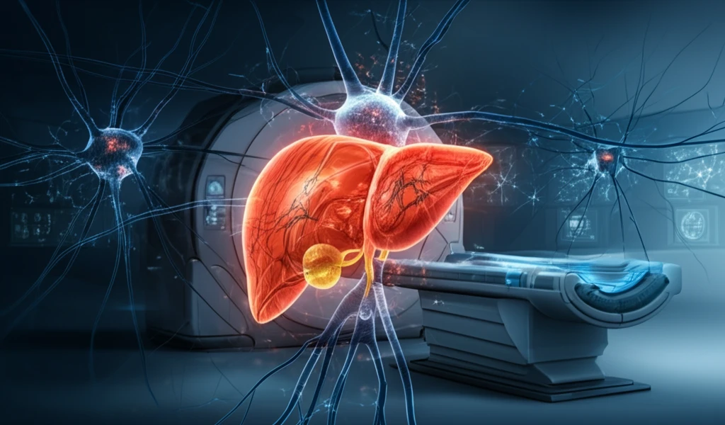
Early Detection of Liver Cancer: A Guide to Advanced Imaging and AI

"Discover how cutting-edge AI-enhanced CT scans are revolutionizing liver cancer detection, offering hope and improving outcomes."
The liver is a vital organ responsible for numerous functions, including protein synthesis and biochemical production essential for digestion. As the only liver you have, its strategic location in the abdominal cavity makes it prone to various diseases. Early detection of liver cancer significantly improves life expectancy and informs treatment strategies.
Traditional methods of liver tumor detection, such as manual segmentation, are time-consuming and can be subjective. To address these limitations, researchers are developing advanced, automated techniques that promise more efficient and accurate diagnoses.
This article explores an innovative method employing Fuzzy C-Means (FCM), Neutrosophic sets (NS), and Bayesian Neural Networks (BNN) to detect cancerous cells in the liver. This approach leverages abdominal CT scans to extract and analyze liver images, providing a more reliable and timely diagnosis.
Liver Cancer Detection: The Scale of the Challenge
Liver cancer remains a leading cause of cancer mortality worldwide, underscoring the need for reliable early detection tools. A comprehensive multi-phase CT dataset, MCT-LTDiag, comprising 517 cases with four-phase contrast-enhanced scans, illustrates the complexity of liver tumor diagnosis and the volume of data needed for robust automated systems. In clinical practice, CT scanning in people with chronic liver disease likely misses liver cancer in approximately 22.5% of patients who would not receive timely treatment, while also producing false-positive results in 8.7% of cases. A large multi-center study analyzing 1,598 contrast-enhanced CT scans from 1,306 cancer patients—identifying 4,908 liver tumors—demonstrates both the prevalence of liver malignancies and the scale at which detection tools must operate.
How CT Scans Detect Liver Cancer—and Where They Fall Short
CT scanning is a primary tool for detecting liver cancer, relying on contrast-enhanced, multi-phase imaging protocols that reveal the distinct blood-flow patterns of malignant tumors. Accurate extraction and segmentation of the liver from CT images remain challenging due to the organ's irregular shape, similar intensity values to adjacent structures, variable contrast levels, and inherent image noise. While specialized contrast-enhanced CT can reveal tumors that a standard scan might miss, the technique's success depends entirely on the imaging protocol used. These limitations motivate ongoing research into pre-processing techniques and AI-driven approaches to improve early detection accuracy.
From X-Rays to Modern CT: A Revolution in Medical Imaging
The history of computed tomography traces back to Wilhelm Conrad Röntgen's discovery of X-ray radiation in 1895 and its rapid adoption in medical diagnostics. As of 2025, the field marks the 50th anniversary of the first CT scan of a live patient—an invention that may represent the greatest revolution in medical imaging since the discovery of x-rays itself. Key historical milestones in CT technology include the earliest days of CT development and the technical steps taken to decrease scan times, both per image and per exam. These advances have transformed CT from a research curiosity into an essential imaging modality used across virtually every area of medicine.
How AI is Revolutionizing Liver Cancer Detection
The core of this innovative approach lies in its methodology, which integrates several advanced techniques to segment and classify tumors in CT images of the liver. The process involves:
- Image Pre-processing: Enhances the quality of CT scan images to reduce noise and improve contrast, ensuring clearer and more defined images for subsequent analysis.
- Image Segmentation: Employs the Fuzzy C-Means (FCM) algorithm combined with Neutrosophic Sets (NS) to accurately delineate the liver and potential tumors from the surrounding tissues. FCM is particularly useful as it allows data points to belong to multiple clusters, reflecting the inherent ambiguity in medical images.
- Feature Extraction: Identifies and extracts key characteristics from the segmented liver images. This includes textural features (assessing the arrangement of pixels) and statistical measures such as entropy, mean, and standard deviation, which help differentiate between healthy and cancerous tissue.
- Classification: Uses a Bayesian Neural Network (BNN) to classify the extracted features and predict whether the cells are cancerous. BNNs are adept at handling uncertainty and can provide probabilistic assessments, offering a more nuanced diagnosis.
AI-Powered Advances in Liver Tumor Detection
Recent research has produced SALSA, a fully automated system for liver tumor segmentation and detection tested on 1,598 contrast-enhanced CT scans from 1,306 patients, identifying 4,908 liver tumors across 1,041 patients. Another study introduces a novel approach integrating 3D U-Net architecture with the Bat Algorithm for hyperparameter optimization, enhancing segmentation accuracy and robustness for automated liver tumor detection in CT images. Conventional segmentation techniques frequently struggle with the complexity of medical images, driving the adoption of sophisticated artificial intelligence methods to improve accuracy. These studies collectively demonstrate that deep learning-based tools can achieve high accuracy in liver tumor detection, reducing the need for manual intervention and enabling radiologists to focus on critical diagnostic decisions.
Evaluating CT Scan Limitations in Liver Cancer Detection
Despite its utility, CT scanning is not infallible for liver cancer detection. In people with chronic liver disease, CT likely misses liver cancer in 22.5% of patients, potentially delaying timely treatment, and produces false-positive results in 8.7% of cases. These limitations highlight that while CT is instrumental in cancer staging—determining how far the cancer has progressed and whether it has spread to lymph nodes or distant organs—its accuracy is contingent on imaging protocol and patient factors. Careful clinical interpretation and complementary imaging modalities remain essential to mitigate the risks of both missed diagnoses and unnecessary interventions.
Comparing Imaging Modalities for Liver Cancer Detection
CT scans can detect small tumors with a sensitivity of approximately 84% for tumors between 10 and 20 mm in size, providing detailed cross-sectional images that aid in staging and treatment planning. Emerging research compares single-energy CT (SECT) with dual-energy CT (DECT) at equivalent radiation doses, assessing whether DECT spectral curve analysis improves detection and characterization accuracy of liver metastases. Ultrasound represents another non-invasive imaging modality used in liver cancer diagnosis, though its comparison with CT involves trade-offs in resolution, accessibility, and operator dependence. Accurate imaging across these modalities is crucial for diagnosing, staging, and planning treatment strategies for primary liver cancer.
The Future of Liver Cancer Diagnostics
The AI-enhanced method demonstrates promising results, achieving an accuracy of 97.33%, sensitivity of 98.03%, and specificity of 97.26%. These metrics indicate the method's high reliability in correctly identifying both cancerous and healthy tissues. With a short time delay of just 3.04 seconds, it also provides a fast diagnostic option. This is a valuable second opinion for doctors and improves patients peace of mind for the accurate diagnosis of their condition at the earliest stages.
Integrating AI Into Clinical Workflows for Liver Diagnosis
The Liver DiagnOsis Network (LiON) represents a significant advance in AI-guided liver malignancy diagnosis, offering a contrast-enhanced CT-based system that supports flexible multiphase processing and clinical data integration. By integrating with existing clinical workflows, such systems aim to provide real-time decision support during liver cancer diagnosis and staging. The development of tools like LiON reflects the growing recognition that AI can augment radiologists' capabilities rather than replace them, supporting more consistent and efficient diagnostic processes. As these AI systems mature, their ability to incorporate multiple imaging phases and clinical data points promises to improve diagnostic accuracy across diverse patient populations.
Emerging Technologies and the Future of Liver Cancer Detection
Advances in deep learning have made it possible to achieve high accuracy in the segmentation and detection of liver tumors from CT scans, eliminating the need for manual intervention and allowing radiologists to focus on critical diagnostic decisions. Ongoing research in epidemiology, risk factors, and diagnostic techniques is essential for improving early diagnosis and reducing the global burden of liver cancer. As most patients are diagnosed at advanced stages, imaging plays a central role across the entire care continuum—from surveillance and early detection to diagnosis, staging, prognostication, and treatment response assessment. The convergence of these technological and clinical advances suggests a future where liver cancer is detected earlier and treated more effectively.
Overcoming Barriers to Equitable Liver Cancer Detection
Despite advances in treatments, obstacles persist in early detection and equity in liver cancer care. Important studies in 2024 highlighted the ongoing global battle against liver cancer, a deadly yet common cancer where disparities in access to advanced imaging and AI-driven diagnostic tools remain significant. The challenge extends beyond technology to include healthcare infrastructure, affordability, and the availability of trained specialists in underserved regions. Addressing these systemic barriers is critical to ensuring that the benefits of advanced imaging and AI reach all patients who need them.
Translating Technology into Better Patient Outcomes
Liver tumors, whether primary or metastatic, significantly impact patient outcomes, making accurate identification and quantification crucial for effective management. Systems like SALSA provide automated tools for precise diagnosis, prognosis, and therapy evaluation, directly translating imaging advances into clinical practice. The ability to automatically detect and characterize liver tumors from CT scans reduces diagnostic delays and supports more personalized treatment strategies. Ultimately, the goal of these technological advances is to improve survival rates and quality of life for patients facing liver cancer.