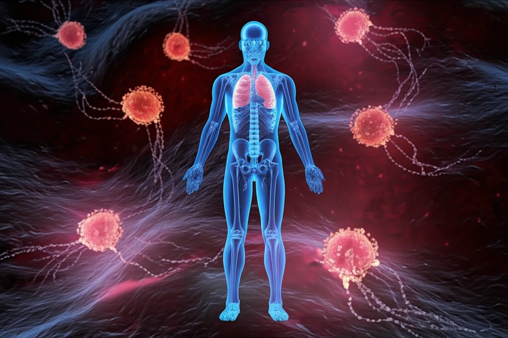
Surreal illustration of glowing nanoparticles detecting cancer cells.

Early Cancer Detection: The Innovative Aptasensor Revolutionizing Diagnostics

"Discover how the new aptasensor technology offers ultrasensitive detection of carcinoembryonic antigen (CEA), paving the way for earlier and more accurate cancer diagnoses."


Cancer remains a leading cause of mortality worldwide, emphasizing the critical need for early and accurate diagnostic tools. Traditional methods often fall short in detecting cancer at its earliest stages, leading to delayed treatment and poorer outcomes. The ability to identify cancer biomarkers with high sensitivity and specificity is crucial for improving survival rates and quality of life for patients.

Carcinoembryonic antigen (CEA) is a well-established biomarker for several types of cancer, including colorectal, pancreatic, and lung cancer. Monitoring CEA levels can provide valuable insights into the presence, progression, and recurrence of these diseases. However, conventional methods for CEA detection may lack the sensitivity required to identify elevated levels in the nascent stages of cancer development.

Recent advancements in biosensor technology have introduced a promising solution: the aptasensor. This innovative device leverages the unique binding properties of aptamers—single-stranded DNA or RNA molecules—to detect specific target substances with remarkable precision. Combining aptamers with cutting-edge techniques like fluorescence resonance energy transfer (FRET) has led to the development of ultrasensitive diagnostic tools capable of detecting CEA at very low concentrations.

The Science Behind the Aptasensor

Surreal illustration of glowing nanoparticles detecting cancer cells.

The aptasensor described in this research article utilizes a sophisticated approach to CEA detection, relying on the principles of fluorescence resonance energy transfer (FRET). This technique involves the transfer of energy between two fluorophores: an energy donor and an energy acceptor. In this case, upconversion nanoparticles (UCNPs) serve as the energy donor, while graphene oxide (GO) acts as the energy acceptor. The magic happens when CEA is introduced to the mix.

The core innovation lies in the design of the aptasensor, which comprises CEA aptamers attached to UCNPs. In the absence of CEA, these aptamer-modified UCNPs bind to GO through π-π stacking interactions. This brings the UCNPs and GO into close proximity, facilitating FRET. As a result, the fluorescence of the UCNPs is quenched, indicating that the aptasensor is in its "off" state. The sensitivity and efficiency of this system makes it an ideal candidate for early cancer detection.

The key components enabling highly accurate cancer biomarker detection are:
  • Upconversion Nanoparticles (UCNPs): Act as energy donors, emitting light upon near-infrared excitation.
  • Graphene Oxide (GO): Functions as an energy acceptor, quenching the fluorescence of UCNPs when in close proximity.
  • CEA Aptamers: Single-stranded DNA or RNA molecules that specifically bind to CEA.
  • Fluorescence Resonance Energy Transfer (FRET): The mechanism by which energy is transferred from UCNPs to GO, resulting in fluorescence quenching.
When CEA is present, the aptamers preferentially bind to the CEA molecules instead of GO. This binding event causes the UCNPs to detach from the GO surface, disrupting FRET. As the UCNPs move away from GO, their fluorescence is restored, signaling the presence of CEA. The intensity of the fluorescence is directly proportional to the concentration of CEA, allowing for quantitative measurement. This method is not only highly sensitive but also selective, ensuring accurate detection even in complex biological samples.

The Future of Cancer Diagnostics

The development of ultrasensitive aptasensors for CEA detection represents a significant step forward in the field of cancer diagnostics. By enabling earlier and more accurate detection of cancer biomarkers, this technology holds the potential to improve patient outcomes and reduce the burden of cancer. Further research and development in this area could lead to the creation of point-of-care diagnostic devices, making cancer screening more accessible and affordable for individuals worldwide. With continued innovation, the aptasensor may soon become an indispensable tool in the fight against cancer.

About this Article -

This article was crafted using a human-AI hybrid and collaborative approach. AI assisted our team with initial drafting, research insights, identifying key questions, and image generation. Our human editors guided topic selection, defined the angle, structured the content, ensured factual accuracy and relevance, refined the tone, and conducted thorough editing to deliver helpful, high-quality information.See our About page for more information.

This article is based on research published under:

DOI-LINK: 10.1016/j.talanta.2018.11.011, Alternate LINK

Title: An Ultrasensitive Homogeneous Aptasensor For Carcinoembryonic Antigen Based On Upconversion Fluorescence Resonance Energy Transfer

Subject: Analytical Chemistry

Journal: Talanta

Publisher: Elsevier BV

Authors: Yujie Wang, Zikai Wei, Xianda Luo, Quan Wan, Rongliang Qiu, Shizhong Wang

Published: 2019-04-01

Everything You Need To Know

1

How does the aptasensor technology detect Carcinoembryonic antigen (CEA)?

The aptasensor works by utilizing CEA aptamers attached to Upconversion Nanoparticles (UCNPs). In the absence of Carcinoembryonic antigen (CEA), these aptamer-modified UCNPs bind to Graphene Oxide (GO), which quenches the fluorescence of the UCNPs through Fluorescence Resonance Energy Transfer (FRET). When Carcinoembryonic antigen (CEA) is present, the CEA aptamers bind to it instead of the Graphene Oxide (GO), restoring the fluorescence of the Upconversion Nanoparticles (UCNPs).

2

What are the key components that enable the aptasensor to accurately detect cancer biomarkers?

The key components enabling highly accurate cancer biomarker detection are: Upconversion Nanoparticles (UCNPs) that act as energy donors, emitting light upon near-infrared excitation; Graphene Oxide (GO) which functions as an energy acceptor, quenching the fluorescence of Upconversion Nanoparticles (UCNPs) when in close proximity; CEA Aptamers that are single-stranded DNA or RNA molecules that specifically bind to Carcinoembryonic antigen (CEA); and Fluorescence Resonance Energy Transfer (FRET) which is the mechanism by which energy is transferred from Upconversion Nanoparticles (UCNPs) to Graphene Oxide (GO), resulting in fluorescence quenching.

3

What advantages does the aptasensor offer over traditional methods of Carcinoembryonic antigen (CEA) detection?

The aptasensor offers a significant advancement because it allows for earlier and more accurate detection of Carcinoembryonic antigen (CEA), a biomarker for several cancers. Traditional methods often lack the sensitivity to detect Carcinoembryonic antigen (CEA) at very low concentrations in the early stages of cancer development. This ultrasensitive detection capability of the aptasensor can lead to earlier diagnosis, timely treatment, and improved patient outcomes. The ability to use Fluorescence Resonance Energy Transfer (FRET) makes the process much more efficient.

4

Can you explain the role of Fluorescence Resonance Energy Transfer (FRET) in the aptasensor's Carcinoembryonic antigen (CEA) detection process?

Fluorescence Resonance Energy Transfer (FRET) is used in the aptasensor for Carcinoembryonic antigen (CEA) detection. Upconversion Nanoparticles (UCNPs) act as energy donors, and Graphene Oxide (GO) acts as the energy acceptor. When Carcinoembryonic antigen (CEA) is absent, Upconversion Nanoparticles (UCNPs) and Graphene Oxide (GO) are in close proximity, causing the Upconversion Nanoparticles (UCNPs) fluorescence to be quenched. When Carcinoembryonic antigen (CEA) is present, the Upconversion Nanoparticles (UCNPs) detach from the Graphene Oxide (GO), restoring fluorescence, indicating Carcinoembryonic antigen (CEA) presence. The implications of using Fluorescence Resonance Energy Transfer (FRET) in this context are far-reaching, as it allows for highly sensitive and specific detection of cancer biomarkers, potentially transforming early cancer diagnostics. The absence of Carcinoembryonic antigen (CEA) allows the process to have a base reading.

5

What are the potential long-term implications of using the aptasensor for cancer diagnostics, and what further research is needed?

The advancement of the aptasensor might result in point-of-care diagnostic devices, which would make cancer screening more accessible and affordable worldwide. Additionally, it could lead to personalized medicine approaches where treatment plans are tailored based on the Carcinoembryonic antigen (CEA) levels detected by the aptasensor. However, further research is needed to validate its effectiveness across diverse populations and cancer types. Understanding the ethics of early screening and potential over-diagnosis is also crucial.

Newsletter Subscribe

Subscribe to get the latest articles and insights directly in your inbox.