Surreal digital illustration of a brain intertwined with DTI metrics.

DTI Scans: A New Window into Glioma Management

"Unlock the potential of Diffusion Tensor Imaging (DTI) for enhanced glioma treatment and outcomes. Learn how DTI's quantitative insights are revolutionizing clinical decisions."


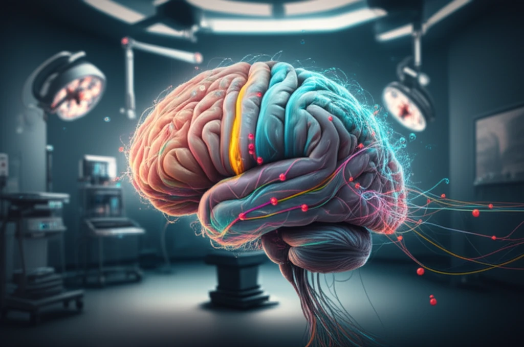
Modern neurosurgery is constantly evolving through technological advancements, with Magnetic Resonance Imaging (MRI) playing a crucial role. Diffusion Tensor Imaging (DTI), a type of MRI, provides a macroscopic view of the microstructures within the brain's white matter. This technique assesses the physiological motion of water in vivo, offering valuable insights for both clinical treatment and glioma research.

The diffusion tensor's mathematical properties enable the extraction of scalar measures, making quantitative analyses of fine-grained imaging characteristics possible. These analyses have the potential to answer many clinical questions, helping to reveal intrinsic details of brain changes under specific pathological circumstances such as glioma.

This article focuses on the merits of quantitative DTI evaluation for glioma management, covering its applications in tissue characterization, white matter tract mapping, radiotherapy delineation, post-therapy outcome assessment, and multimodal imaging. A presentation of DTI's limitations is also elucidated.

DTI Metrics: Guiding Precision in Glioma Treatment

Surreal digital illustration of a brain intertwined with DTI metrics.

DTI metrics are essential for characterizing tissue, mapping white matter tracts, and planning treatments. Fractional anisotropy (FA) measures the directionality of water diffusion, ranging from 0 (isotropic) to 1 (anisotropic), while mean diffusivity (MD) measures the magnitude of diffusion. MD is mathematically equivalent to the apparent diffusion coefficient (ADC) in isotropic diffusion-weighted imaging (DWI).

While valuable, the quantitative measurement of DTI metrics is influenced by several factors that must be considered. These include:

  • Cross-sectional variability of patients
  • MRI scanner parameters
  • Voxel size
  • ROI definition
  • Image acquisition artifacts
  • Data pre-processing tools
  • Tensor fitting algorithm
  • Free water content
  • Tumor pathology and heterogeneity
  • Fiber tract alterations due to tumor invasion
  • Interstitial free water accumulation
  • Patient treatment regime
Corrected DTI yields increased precision and a more complete tract reconstruction. Factors influencing DTI measures need to be standardized during both acquisition and post-processing. Physicians must understand the limitations and potential pitfalls of this technique when making DTI-based clinical decisions in glioma management.

Maximizing DTI's Potential in Neurosurgery

DTI metrics evaluation and tractography offer promise as a non-invasive brain mapping technique in neurosurgery for diagnosis, surgical planning, and post-therapy assessment.

As DTI becomes more widely used in clinical and research settings, radiologists, medical physicists, neuroscientists, and biomedical engineers will play essential roles alongside neurosurgeons to overcome its limitations.

By working together, these experts can continually improve and provide the best medical care to patients, pushing the boundaries of what's possible in glioma management.

About this Article -

This article was crafted using a human-AI hybrid and collaborative approach. AI assisted our team with initial drafting, research insights, identifying key questions, and image generation. Our human editors guided topic selection, defined the angle, structured the content, ensured factual accuracy and relevance, refined the tone, and conducted thorough editing to deliver helpful, high-quality information.See our About page for more information.

This article is based on research published under:

DOI-LINK: 10.1007/s10143-018-1050-1, Alternate LINK

Title: Quantitative Evaluation Of Diffusion Tensor Imaging For Clinical Management Of Glioma

Subject: Neurology (clinical)

Journal: Neurosurgical Review

Publisher: Springer Science and Business Media LLC

Authors: Ye Li, Wenyao Zhang

Published: 2018-11-12

Everything You Need To Know

1

What exactly is Diffusion Tensor Imaging (DTI) and how does it offer insights into glioma?

Diffusion Tensor Imaging (DTI) is a type of Magnetic Resonance Imaging (MRI) that provides a detailed view of the brain's white matter microstructures by assessing the physiological motion of water. This allows for a quantitative analysis using scalar measures to reveal intrinsic details of brain changes, particularly in pathological conditions such as glioma.

2

What factors can influence the measurements obtained from Diffusion Tensor Imaging (DTI), and why is standardization important?

Diffusion Tensor Imaging (DTI) metrics are affected by several factors, including cross-sectional variability of patients, MRI scanner parameters, voxel size, ROI definition, image acquisition artifacts, data pre-processing tools, tensor fitting algorithm, free water content, tumor pathology and heterogeneity, fiber tract alterations due to tumor invasion, interstitial free water accumulation and patient treatment regime. Standardization of these factors during both acquisition and post-processing is crucial for increased precision and complete tract reconstruction.

3

Can you explain the difference between Fractional anisotropy (FA) and Mean diffusivity (MD) in Diffusion Tensor Imaging (DTI)?

Fractional anisotropy (FA) measures the directionality of water diffusion, ranging from 0 (isotropic) to 1 (anisotropic). Mean diffusivity (MD) measures the magnitude of water diffusion. MD is mathematically equivalent to the apparent diffusion coefficient (ADC) in isotropic diffusion-weighted imaging (DWI).

4

In what specific ways is quantitative Diffusion Tensor Imaging (DTI) being used in the management of glioma?

Quantitative Diffusion Tensor Imaging (DTI) is used in glioma management for tissue characterization, white matter tract mapping, radiotherapy delineation, post-therapy outcome assessment, and multimodal imaging. It helps in understanding the intricate details of brain changes under specific pathological circumstances.

5

What are the limitations of Diffusion Tensor Imaging (DTI) and how can physicians ensure accuracy when using DTI-based clinical decisions in glioma management?

While Diffusion Tensor Imaging (DTI) offers valuable insights, it's essential for physicians to understand its limitations and potential pitfalls when making DTI-based clinical decisions in glioma management. Corrected DTI yields increased precision and a more complete tract reconstruction when the different factors that influence DTI measures are standardized during both acquisition and post-processing. It is a non-invasive brain mapping technique in neurosurgery for diagnosis, surgical planning, and post-therapy assessment.

Newsletter Subscribe

Subscribe to get the latest articles and insights directly in your inbox.