Digoxin's Deceptive Dance: When Heart Medication Mimics a Heart Attack
"Learn how digoxin, a common heart medication, can sometimes produce ECG patterns that mimic critical heart ischemia, leading to unnecessary alarm and interventions."
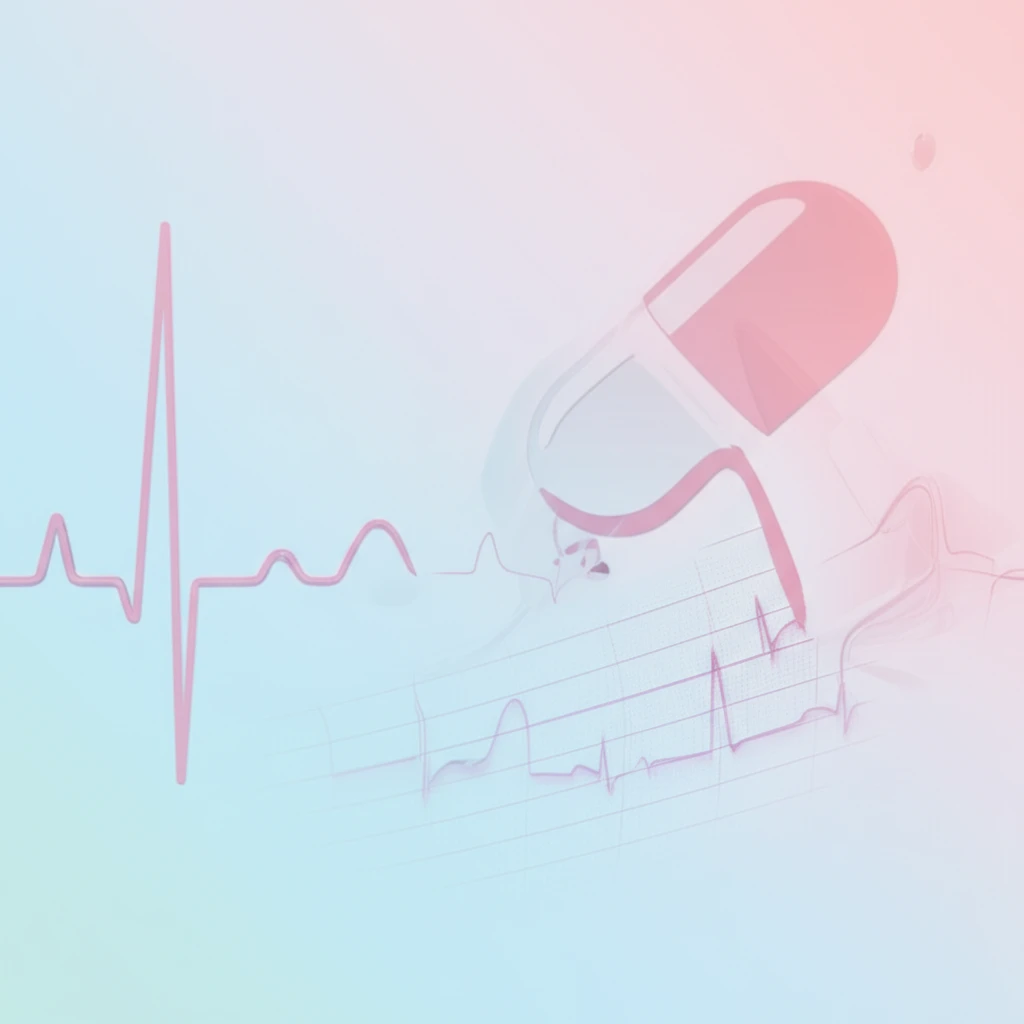
In the world of cardiology, accurately interpreting an electrocardiogram (ECG) is paramount. An ECG provides a snapshot of the heart's electrical activity, helping doctors diagnose various conditions, including myocardial ischemia (reduced blood flow to the heart) and heart attacks. However, certain medications can sometimes muddy the waters, creating ECG patterns that mimic these dangerous conditions.
One such medication is digoxin, a drug commonly prescribed for atrial fibrillation (an irregular heartbeat) and heart failure. While digoxin can be a life-saving treatment, it's also known to cause distinctive changes on an ECG. The challenge arises when these digoxin-related ECG changes resemble those seen in critical myocardial ischemia, potentially leading to misdiagnosis and unnecessary interventions.
This article delves into a fascinating case where a patient on digoxin experienced chest pain, and their ECG showed patterns strikingly similar to those of a heart attack. We'll explore how doctors navigated this diagnostic puzzle, ultimately uncovering that digoxin, not a blocked artery, was the culprit. This case underscores the importance of considering medication effects when interpreting ECGs and highlights the potential for misdiagnosis, even in critical situations.
Decoding Digoxin's ECG Deception: What to Look For
Digoxin affects the heart's electrical activity, leading to characteristic changes on an ECG. These changes can include ST-segment depression (a dip in a specific part of the ECG waveform) and T-wave abnormalities. While these changes are often benign, they can sometimes resemble the patterns seen in myocardial ischemia, particularly in specific leads (ECG recording points).
- Clinical Context: Has the patient been taking Digoxin? What are the other medications? Any recent changes in Dosage?
- ECG Morphology: Digoxin typically causes a gradual, "scooped" ST-segment depression, while ischemia often produces a more angular or horizontal ST-segment depression.
- Reciprocal Changes: Ischemia often produces reciprocal changes (mirror-image patterns) in other ECG leads, which are less common with digoxin effect.
- Digoxin Level: Measuring the digoxin level in the blood can help determine if the patient is within the therapeutic range or experiencing toxicity. However, ECG changes can occur even with normal digoxin levels.
- Clinical Presentation: The overall clinical picture, including the patient's symptoms and other medical history, is crucial in making the correct diagnosis.
The Takeaway: Vigilance and Careful Interpretation
This case serves as a powerful reminder that accurate ECG interpretation requires a holistic approach. While ECGs are invaluable diagnostic tools, they should always be interpreted in the context of the patient's clinical history, medication list, and other relevant factors. When digoxin is involved, clinicians must be particularly vigilant in differentiating between drug-induced ECG changes and those indicative of true myocardial ischemia. This careful approach can help avoid unnecessary interventions and ensure patients receive the most appropriate and timely care.