Dermoscopy aiding in splinter removal

Dermoscopy: The Unexpected Key to Finding Hidden Splinters

"Learn how dermoscopy, a technique typically used for skin exams, can help doctors quickly and easily locate and remove subungual foreign bodies like splinters and fish bones."


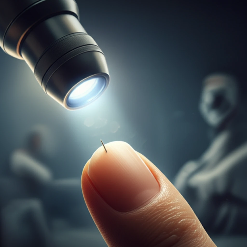
Finding a splinter can be a minor nuisance, but when it's hidden deep under a fingernail, it can become a frustrating ordeal. Traditional methods often fall short, leaving you poking and prodding with limited success. But what if there was a better way? A technique usually reserved for examining moles and skin conditions might just be the answer: dermoscopy.

Dermoscopy is a non-invasive imaging technique that uses a special magnifying lens and light source to visualize the skin in greater detail. While it's primarily used by dermatologists to diagnose skin cancer and other skin disorders, its potential extends beyond the surface. Recent studies have shown that dermoscopy can also be a valuable tool for identifying and removing foreign bodies lodged beneath the nails – a condition known as subungual foreign bodies.

A recent report highlighted a case where dermoscopy successfully aided in the identification and removal of a fish bone embedded under a woman's fingernail. This case underscores the potential of dermoscopy as a quick, effective, and minimally invasive method for dealing with those pesky hidden splinters and other foreign objects that find their way under our nails.

The Case for Dermoscopy: A Clearer View of Hidden Objects

Dermoscopy aiding in splinter removal

The case report, published in the journal Annals of Dermatology, details the experience of a 51-year-old woman who sought medical help after struggling to remove a fish bone splinter from her thumb. Despite her best efforts, she couldn't locate the foreign body, leading to persistent pain and discomfort.

Upon examination, doctors found it difficult to pinpoint the exact location of the fish bone with the naked eye. That's when they turned to dermoscopy. The dermoscopic examination revealed a clear image of a linear foreign body embedded under the nail plate. With the aid of this magnified view, the doctors were able to easily remove the fish bone using fine forceps.

Here's why dermoscopy is proving to be so useful in these situations:
  • Enhanced Visualization: Dermoscopy provides a magnified and illuminated view of the area under the nail, making it easier to identify foreign bodies that are otherwise difficult to see.
  • Non-Invasive: The procedure is entirely non-invasive, causing no pain or trauma to the patient.
  • Quick and Efficient: Dermoscopy can be performed quickly and easily in a clinical setting, allowing for prompt diagnosis and treatment.
  • Improved Accuracy: By providing a clearer view of the foreign body, dermoscopy helps to ensure complete removal, reducing the risk of complications such as infection.
Traditional methods of foreign body removal often involve blind probing or even surgical exploration, which can be painful and potentially damaging to the nail and surrounding tissues. Dermoscopy offers a gentler, more precise alternative, minimizing discomfort and improving outcomes.

A Promising Tool for Everyday Ailments

While dermoscopy is primarily used in dermatology, its application in identifying and removing subungual foreign bodies highlights its versatility as a diagnostic tool. For those frustrating moments when you can't seem to locate that tiny splinter, dermoscopy might just be the answer. Talk to your doctor or a dermatologist about whether dermoscopy could be a helpful option for you.

About this Article -

This article was crafted using a human-AI hybrid and collaborative approach. AI assisted our team with initial drafting, research insights, identifying key questions, and image generation. Our human editors guided topic selection, defined the angle, structured the content, ensured factual accuracy and relevance, refined the tone, and conducted thorough editing to deliver helpful, high-quality information.See our About page for more information.

This article is based on research published under:

DOI-LINK: 10.5021/ad.2016.28.1.144, Alternate LINK

Title: The Usefulness Of Dermoscopy For Detection Of Subungual White Foreign Bodies

Subject: Dermatology

Journal: Annals of Dermatology

Publisher: Korean Dermatological Association and The Korean Society for Investigative Dermatology

Authors: Hyang-Suk You, Gun-Wook Kim, Won-Jeong Kim, Je-Ho Mun, Margaret Song, Hoon-Soo Kim, Hyun-Chang Ko, Byung-Soo Kim, Moon-Bum Kim

Published: 2016-01-01

Everything You Need To Know

1

What is dermoscopy and how is it typically used?

Dermoscopy is a non-invasive skin examination technique that uses a special magnifying lens and light source to visualize the skin in greater detail. It is primarily used by dermatologists to diagnose skin cancer and other skin disorders. However, its application extends beyond dermatology, as shown by its ability to help locate subungual foreign bodies.

2

How does dermoscopy aid in the removal of subungual foreign bodies like splinters?

Dermoscopy enhances the visualization of the area under the nail, providing a magnified and illuminated view that makes it easier to identify foreign bodies, which would otherwise be difficult to see. The use of dermoscopy in identifying subungual foreign bodies is non-invasive, quick, efficient and improves the accuracy of foreign body identification.

3

What are the advantages of using dermoscopy over traditional methods for removing splinters hidden under the nail?

Traditional methods of foreign body removal often involve blind probing or even surgical exploration, which can be painful and potentially damaging to the nail and surrounding tissues. Dermoscopy offers a gentler, more precise alternative, minimizing discomfort and improving outcomes due to enhanced visualization, accuracy, and its non-invasive nature.

4

Can you describe a specific instance where dermoscopy proved useful in locating a foreign object?

A case report published in the journal *Annals of Dermatology* detailed the experience of a 51-year-old woman with a fish bone splinter under her thumb. Doctors used dermoscopy to reveal a clear image of the linear foreign body embedded under the nail plate and with the aid of this magnified view, the doctors were able to easily remove the fish bone using fine forceps.

5

Beyond finding splinters, what are the broader implications of using dermoscopy as a diagnostic tool?

While dermoscopy is primarily used in dermatology for diagnosing skin conditions, its successful application in identifying and removing subungual foreign bodies highlights its versatility as a diagnostic tool. This suggests that dermoscopy could potentially be applied in other areas of medicine where enhanced visualization of small or hidden structures is needed, expanding its role beyond traditional dermatological uses. Future research might explore its utility in identifying other types of foreign bodies or in guiding minimally invasive procedures.

Newsletter Subscribe

Subscribe to get the latest articles and insights directly in your inbox.