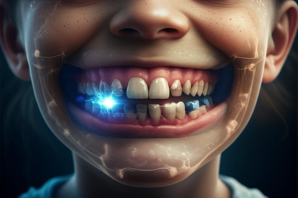
A digital illustration showing a child's face with an x-ray effect, highlighting the advanced technology used in dental tomography.

Decoding Your Smile: How Dental Tomography is Revolutionizing Mixed-Dentition Analysis

"Unlocking the Secrets of Your Child's Smile: Exploring the Cutting-Edge World of Dental Tomography and Its Impact on Orthodontic Treatment."


The journey to a perfect smile often begins in childhood, a time when a child's mouth undergoes significant transformations. For parents, understanding this process and ensuring proper dental development can be a source of both excitement and concern. Mixed-dentition analysis, the assessment of both baby and permanent teeth, is a crucial step in this journey, guiding orthodontists in making informed decisions about future treatments.

Traditional methods for analyzing mixed dentition, such as X-rays and plaster cast models, have long been the standard. However, recent advancements in technology, particularly the use of Cone-Beam Computed Tomography (CBCT), are offering a new level of precision and insight. This innovative technique provides a three-dimensional view of the teeth and jaw, enabling orthodontists to make more accurate predictions and diagnoses.

This article delves into the world of dental tomography, exploring how it compares to traditional methods and the benefits it offers for your child's orthodontic future. We'll explore the advancements in this field, what it means for you, and the impact these innovations have on the future of smiles.

Tomography vs. Traditional Methods: A Comparative Analysis

A digital illustration showing a child's face with an x-ray effect, highlighting the advanced technology used in dental tomography.

Traditional methods for assessing mixed dentition involve a combination of techniques, each with its own set of limitations. X-rays, for example, provide a two-dimensional view, which can make it challenging to visualize the full picture. Plaster cast models offer a physical representation of the teeth but lack the ability to see what lies beneath the gums. These techniques have served orthodontists for decades, but they also come with challenges.

Cone-Beam Computed Tomography (CBCT) represents a leap forward in dental imaging. Unlike traditional X-rays, CBCT provides a three-dimensional view, offering a comprehensive assessment of the teeth, jaw, and surrounding structures. This advanced technology allows orthodontists to see the size, position, and angulation of teeth, including those that have not yet erupted. This level of detail is invaluable for predicting future dental development and planning orthodontic treatment.

  • Enhanced Visualization: CBCT provides a 3D view, allowing for a more comprehensive understanding of dental structures.
  • Improved Accuracy: CBCT offers more precise measurements, reducing the guesswork in treatment planning.
  • Reduced Radiation: Modern CBCT devices emit lower radiation doses compared to traditional CT scans.
  • Predictive Capabilities: CBCT aids in predicting the eruption of teeth and potential future problems.
  • Personalized Treatment: CBCT data facilitates the creation of customized treatment plans.
The key difference lies in the level of detail and the ability to visualize the teeth and jaw in three dimensions. While traditional methods offer valuable insights, CBCT elevates the diagnostic process, leading to better treatment outcomes and a more informed approach to orthodontic care.

The Future of Orthodontics is Now

Dental tomography is reshaping the landscape of orthodontics, offering a new era of precision, accuracy, and personalized care. As technology continues to advance, we can anticipate even more sophisticated imaging techniques that will further enhance our ability to predict, diagnose, and treat dental issues. By understanding these advancements, you can actively participate in the journey toward a healthy, beautiful smile for your child. The future is bright, and the smiles ahead will be even more radiant.

About this Article -

This article was crafted using a human-AI hybrid and collaborative approach. AI assisted our team with initial drafting, research insights, identifying key questions, and image generation. Our human editors guided topic selection, defined the angle, structured the content, ensured factual accuracy and relevance, refined the tone, and conducted thorough editing to deliver helpful, high-quality information.See our About page for more information.

Everything You Need To Know

1

What is mixed-dentition analysis and why is it important for my child?

Mixed-dentition analysis is the assessment of both baby teeth and permanent teeth in a child's mouth. This analysis is a crucial step because it guides orthodontists in making informed decisions about future orthodontic treatments. It helps in understanding the development of your child's smile and addressing potential issues early on.

2

How does Cone-Beam Computed Tomography (CBCT) enhance orthodontic treatment planning compared to traditional X-rays?

Cone-Beam Computed Tomography (CBCT) offers a three-dimensional view of the teeth, jaw, and surrounding structures, unlike traditional X-rays, which only provide a two-dimensional view. This allows orthodontists to see the size, position, and angulation of teeth, including those that have not yet erupted, leading to more accurate predictions, diagnoses, and personalized treatment plans. Traditional X-rays can be limiting as they don't provide the comprehensive assessment that CBCT does.

3

What are the benefits of using Cone-Beam Computed Tomography (CBCT) in my child's orthodontic assessment?

Cone-Beam Computed Tomography (CBCT) offers several benefits including enhanced visualization through its 3D view, improved accuracy in measurements which reduces guesswork in treatment planning, reduced radiation doses compared to traditional CT scans, predictive capabilities for tooth eruption and potential future problems, and facilitation of personalized treatment plans based on detailed data.

4

In what ways does dental tomography with Cone-Beam Computed Tomography (CBCT) improve the accuracy of orthodontic predictions and diagnoses, and what impact does this have on treatment outcomes?

Dental tomography using Cone-Beam Computed Tomography (CBCT) provides a comprehensive, three-dimensional view of the teeth, jaw, and surrounding structures. This allows orthodontists to assess the size, position, and angulation of teeth with greater precision, even for unerupted teeth. This enhanced accuracy in predictions and diagnoses leads to more effective and personalized treatment plans, ultimately improving treatment outcomes and ensuring a healthier, more beautiful smile.

5

How is Cone-Beam Computed Tomography (CBCT) contributing to the future of orthodontics and personalized dental care for children?

Cone-Beam Computed Tomography (CBCT) is revolutionizing orthodontics by offering unprecedented precision, accuracy, and personalized care. The detailed 3D imaging allows for better prediction, diagnosis, and treatment of dental issues. As technology advances, more sophisticated imaging techniques are expected to further enhance the ability to provide tailored orthodontic solutions, ensuring healthier and more radiant smiles for children. This represents a shift toward proactive and individualized dental care, optimizing treatment outcomes and patient satisfaction.

Newsletter Subscribe

Subscribe to get the latest articles and insights directly in your inbox.