Decoding the Shadows: Unraveling the Mysteries of Breast Cancer Risk and Mammographic Density
"Understanding the Link: How Mammographic Density and Breast Cancer Risk Intertwine"
Breast cancer remains a significant health concern for women worldwide. Early detection is key, and mammography is a crucial tool in this fight. However, the interpretation of mammograms, particularly regarding mammographic density, can be complex, leading to questions about its role in risk assessment and screening strategies.
This article delves into the latest research exploring the relationship between mammographic density and breast cancer risk. We'll break down the science behind density, discuss the challenges in using it as a standalone risk factor, and shed light on how this information impacts your breast health journey.
We will explore the findings from studies, including those published in the journal Radiology, providing a clear understanding of the strengths and limitations of various density measurement methods.
Unpacking Mammographic Density: What It Is and Why It Matters
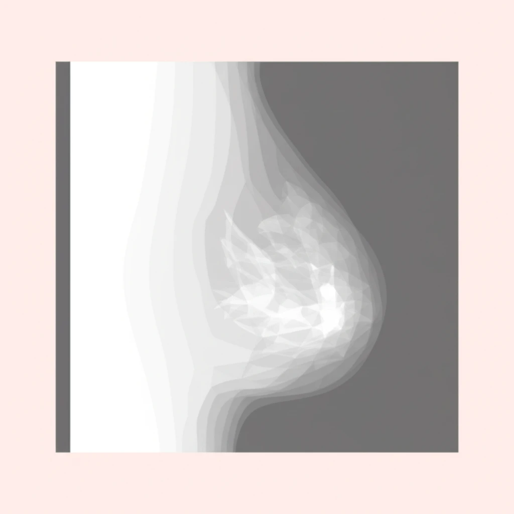
Mammographic density refers to the proportion of fibrous and glandular tissue compared to fatty tissue within the breast. This density appears as white areas on a mammogram, while fatty tissue appears dark. Higher density can make it more challenging to detect tumors, as both dense tissue and tumors appear white. Also, increased density is a known risk factor for breast cancer.
- BI-RADS: A widely used system that categorizes density into four categories, from almost entirely fatty (A) to extremely dense (D).
- Automated Methods: These include methods like Cumulus and Volpara, which use computer algorithms to quantify density.
Empowering Your Breast Health: Moving Forward
Understanding mammographic density is an essential step in proactive breast health management. This knowledge enables you to have informed conversations with your healthcare providers, participate in personalized screening plans, and advocate for your well-being. Remember, you are not alone, and there are resources available to guide you on your journey to optimal breast health.